Upload presentasi
Presentasi sedang didownload. Silahkan tunggu

1
CURRICULUM VITAE NAMA : dr. NASDALDY, Sp.OG
TEMPAT & TGL. LAHIR : BUKIT TINGGI, 8 – 12 – 1955 ALAMAT KANTOR : JL. S. PARMAN KAV. 84 – 86, SLIPI JAKARTA BARAT TELP EXT. 2501 ALAMAT RUMAH : JL. KWENI NO. 17, GANDARIA UTARA JAKARTA SELATAN PENDIDIKAN TERAKHIR : SPESIALIS OBSTETRI GINEKOLOGI DI FKUI TAHUN 1988 PEKERJAAN SAAT INI : KETUA KELOMPOK STAF MEDIK FUNGSIONAL RSK. DHARMAIS

2
KANKER SERVIKS SKRINING, DIAGNOSA DAN PENATALAKSANAAN
dr. Nasdaldy, Sp.OG Divisi Kanker Ginekologi RS. Kanker “Dharmais” Jakarta

3
Apa itu Kanker Serviks ? Kanker Leher Rahim :
Adalah kanker yang terjadi pada leher rahim (serviks) 1 Apa itu serviks ? Serviks adalah daerah yang menghubungkan rahim (uterus) dan vagina. GCF (Gynecologic Cancer Foundation) Presentation The Adam Health Illustrated Encyclopedia, A.D.A.M., Inc. is accredited by URAC, also known as the American Accreditation HealthCare Commission
 1. Apa itu serviks Serviks adalah daerah yang menghubungkan rahim (uterus) dan vagina. GCF (Gynecologic Cancer Foundation) Presentation. The Adam Health Illustrated Encyclopedia, A.D.A.M., Inc. is accredited by URAC, also known as the American Accreditation HealthCare Commission.")
4
Organ Genitalia Perempuan
Leher rahim

5
Seberapa sering Kanker Serviks ?
Menurut WHO TIAP TAHUN, DI SELURUH DUNIA: 490,000 perempuan didiagnosa* menderita kanker serviks 240,000 di antaranya MENINGGAL *80% terjadi di negara berkembang

6
Banyaknya Kasus dan Kematian dari Kanker Serviks
Prediksi Kasus dan Kematian Kanker Serviks di tahun 2002 14, United States/ Canada 59,929 29,814 Europe 61,132 31,314 Eastern Asia 157,759 86,708 Southcentral Asia 17, Central America 42,538 22,594 Southeast Asia 78,896 61,670 Africa 48,328 21,402 South America 1, Australia/ New Zealand 1. Ferlay J, Bray F, Pisani P, Parkin DM. Lyon, France: IARC Press; 2004.

7
Bagaimana Terjadinya Kanker Serviks?
Kanker serviks dapat berkembang ketika sel yang abnormal dalam serviks mulai membelah diri tanpa terkendali Sel yang abnormal pada serviks dapat berkumpul menjadi tumor Jinak tidak berbahaya tetap pada daerah sumbernya, tidak menyebar TUMOR Ganas berbahaya , dapat menjadi kanker akan menyebar ke daerah lain The Adam Health Illustrated Encyclopedia, A.D.A.M., Inc. is accredited by URAC, also known as the American Accreditation HealthCare Commission

8
Bagaimana terjadinya Kanker Serviks ?
Sel Normal Lesi Pra Kanker Kanker

9
Cervical Cancer

10
Serviks Normal (Leher Rahim)
")
11
Lesi Pra Kanker 1/Wright/p.2120/abstract conclusions 2/Bonnez/p. 576/Figure 12; p. 578/col 1/¶2 3/CCS/p 6/¶2 4/Sellors/Ch. 7/p.9/Figure 7.23. 1. Wright TC Jr, Cox JT, Massad LS, et al, for the ASCCP-Sponsored Consensus Congress. JAMA. 2002;287:2120– Bonnez W. In: Richman DD, Whitley RJ, Hayden FJ, eds. Washington, DC: American Society for Microbiology Press; 2002:557– Canadian Cancer Society. Cervical Cancer: What you need to know. Available at: Accessed March 13, Reprinted with permission from Sellors JW, Sankaranarayanan R, eds. Colposcopy and Treatment of Cervical Intraepithelial Neoplasia. A Beginner’s Manual. Lyon, France: International Agency for Research on Cancer; 2003. 1/Sellors/Ch. 4/p. 1/¶ 1; p.5/¶ 2 1/Sellors/Ch. 7/p. 1/¶ 1; p. 6/¶ 3, Figure 7.12. 1/Sellors/Ch. 7/p. 1/¶ 1; p. 6/¶3,4; p. 8/ Figure 7.20; p.9/Figure 7.23.

12
Apa penyebab Kanker Serviks?
KANKER SERVIKS DISEBABKAN OLEH HUMAN PAPILLOMAVIRUSa,1,2 120 Tipe HPV telah diketahui 30-40 Tipe HPV menyerang anogenital Low risk type ( HPV 6 & 11 ) (tidak menyebabkan kanker) Menyebakan anogenital warts High risk type ( HPV 16 & 18) Menyebabkan kanker serviks Anogenital : area kelamin (termasuk kulit penis, mulut vagina & anus) Infeksi dengan HPV seringkali TIDAK menimbulkan gejala Banyak orang TIDAK tidak tahu mereka terinfeksi HPV Banyak orang dapat menularkan HPV TANPA menyadarinya
 (tidak menyebabkan kanker) Menyebakan anogenital warts. High risk type ( HPV 16 & 18) Menyebabkan kanker serviks. Anogenital : area kelamin (termasuk kulit penis, mulut vagina & anus) Infeksi dengan HPV seringkali TIDAK menimbulkan gejala. Banyak orang TIDAK tidak tahu mereka terinfeksi HPV. Banyak orang dapat menularkan HPV TANPA menyadarinya.")
13
Cara Penularan

14
Pap Smears sebagai Deteksi Dini
1/Parham/p. S14/Table 1. 2/Schink/p. 5/ col 2/¶2. 3/Uyar/p. 227/ abstract. 4/Kulasingam/p. 1754/Table 3. Pap Smear : pengambilan sel dari serviks, diperiksa dengan mikroskop untuk mengetahui adanya kelainan pada serviks 2/Schink/p. 5/col 2/¶1. 5/Selvaggi/ p. 1506/col 1/¶1, 3. 6/Chacho/p. 137/col 2/¶5 1/Hewitt/p. 270/ col 1/¶1 1. Hewitt M, Devesa SS, Breen N. Cervical cancer screening among U.S. women: analyses of the 2000 National Health Interview Survey. Prev Med. 2004;39: 2. Crum CP, Rivera MN. Vaccines for cervical cancer. Cancer J. 2003;9:368–376. 3. Sung HY, Kearney KA, Miller M, Kinney W, Sawaya GF, Hiatt RA. Papanicolaou smear history and diagnosis of invasive cervical carcinoma among members of a large prepaid health plan. Cancer. 2000;88:2283–2289. 4. Schink JC. Strategies for detecting cervical dysplasia: Visual inspection, spectroscopy, and speculoscopy. OBG Manag. 2003;(suppl):5–8. 5. Selvaggi SM. Implications of low diagnostic reproducibility of cervical cytologic and histologic diagnoses. JAMA. 2001;285:1506–1508. 6. Chacho MS, Mattie ME, Schwartz PE. Cytohistologic correlation rates between conventional Papanicolaou smears and ThinPrep cervical cytology: A comparison. Cancer. 2003;99:135–140. 7. Saslow D, Runowicz CD, Solomon D, et al. American Cancer Society Guideline for the early detection of cervical neoplasia and cancer. CA Cancer J Clin. 2002;52:342–362. 8. Uyar DS, Eltabbakh GH, Mount SL. Positive predictive value of liquid-based and conventional cervical Papanicolaou smears reported as malignant. Gynecol Oncol. 2003;89:227–232. 9. Kulasingam SL, Hughes JP, Kiviat NB, et al. Evaluation of human papillomavirus testing in primary screening for cervical abnormalities: Comparison of sensitivity, specificity, and frequency of referral. JAMA. 2002;288:1749–1757. 2/Crum/p. 368/col 1/¶2; col 2/¶1 3/Sung/p. 2285/col 2/¶3, 4 JIKA ANDA SUDAH MENIKAH / MELAKUKAN HUBUNGAN, LAKUKAN PAP SMEAR SECARA TERATUR 4/Schink/ p. 5/ col 2/¶1. 5/Selvaggi/ p. 1506/col 1/¶1, 3. 6/Chacho/ p. 137/col 2/¶5 5/Selvaggi/ p. 1506/col 1/¶3 7/Saslow/p. 352/col 2/¶2. 8/Uyar/ p. 227/abstract 9/Kulasingam / p.1754/Table 3; p. 1749/abstract 8/Uyar/p. 227/abstract 5/Selvaggi/ p. 1506/col 2/¶1.
:5–8. 5. Selvaggi SM. Implications of low diagnostic reproducibility of cervical cytologic and histologic diagnoses. JAMA. 2001;285:1506– Chacho MS, Mattie ME, Schwartz PE. Cytohistologic correlation rates between conventional Papanicolaou smears and ThinPrep cervical cytology: A comparison. Cancer. 2003;99:135– Saslow D, Runowicz CD, Solomon D, et al. American Cancer Society Guideline for the early detection of cervical neoplasia and cancer. CA Cancer J Clin. 2002;52:342– Uyar DS, Eltabbakh GH, Mount SL. Positive predictive value of liquid-based and conventional cervical Papanicolaou smears reported as malignant. Gynecol Oncol. 2003;89:227– Kulasingam SL, Hughes JP, Kiviat NB, et al. Evaluation of human papillomavirus testing in primary screening for cervical abnormalities: Comparison of sensitivity, specificity, and frequency of referral. JAMA. 2002;288:1749– /Crum/p. 368/col 1/¶2; col 2/¶1. 3/Sung/p. 2285/col 2/¶3, 4. JIKA ANDA SUDAH MENIKAH / MELAKUKAN HUBUNGAN, LAKUKAN PAP SMEAR SECARA TERATUR. 4/Schink/ p. 5/ col 2/¶1. 5/Selvaggi/ p. 1506/col 1/¶1, 3. 6/Chacho/ p. 137/col 2/¶5. 5/Selvaggi/ p. 1506/col 1/¶3. 7/Saslow/p. 352/col 2/¶2. 8/Uyar/ p. 227/abstract. 9/Kulasingam / p.1754/Table 3; p. 1749/abstract. 8/Uyar/p. 227/abstract. 5/Selvaggi/ p. 1506/col 2/¶1.")
15
Cervical Cancer Screening
Pap Smears Regular Pap smears untuk setiap wanita seksual aktif berapapun usianya. Batas umur ????

16
Penting untuk di-INGAT!!!
Kanker serviks adalah kanker yang banyak menyebabkan kematian pada perempuan Kanker serviks dapat dicegah : Deteksi sedini mungkin dengan PAP SMEAR Edukasi mengenai kanker serviks Vaksinasi HPV VAKSIN American Cancer Detailed Guide. American Cancer Society Online Publication. KONSULTASIKAN KEPADA DOKTER ANDA TENTANG KANKER SERVIKS & CARA PENCEGAHANNYA

17
CIN 2/3 dan AIS Penanda yang mewakili untuk Kanker serviks
Definitive Efficacy 0 - 1 tahun 0 - 5 tahun sampai 20 tahun Infeksi awal HPV Infeksi Persistent CIN 2/3 or AIS Kanker serviks CIN 1 The next issue we wanted to address was how to make sure that we get a cancer prevention indication at launch. Such a claim will be critical to the success of the vaccine. Well, lets go back to the schematic of HPV infection natural history in women. Cervical cancer is all the way on the right here. And if you add up all the years between initial HPV infection and cancer, you get to 20 years or so. Too long for a clinical trial. Also, we couldn’t allow women to remain untreated and to develop cancer in a clinical trial – trial participants would have to get the very best health care during the study – including intensive pap screening. So we couldn’t ethically let them progress to cancer. So if cervical cancer is not a feasible endpoint for a clinical trial, how do we get a cervical cancer prevention claim? Well, we looked at the natural history of HPV disease, and asked: what is the very best surrogate marker for cervical cancer? Which pre-cancer lesion is closely tied to cervical cancer that if we show a reduction in this endpoint, we would be very sure that we will see a reduction in cancer? The answer was CIN 2/3, or high grade dysplasia Why not another lesion? Infeksi HPV “sembuh”

18
Vaksin HPV diberikan kepada siapa ?????
Pria usia 9 – 15 tahun Pria usia diatas 15 tahun ??? (On Going Study) Wanita usia 9 – 26 tahun Wanita usia diatas 26 tahun ????
 Wanita usia 9 – 26 tahun. Wanita usia diatas 26 tahun")
19
Bagaimana mendiagnosisnya ?
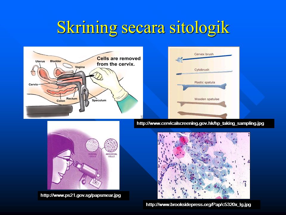
Pap smear: Pengambilan sel – sel serviks dengan spatula atau sitobrush, kemudian dioleskan di objek glas. Setelah itu diperiksa dengan mikroskop. Pelvic examination: Vagina dan organ sekitar diperiksa secara visual dan bimanual (menggunakan dua tangan). Kemudian organ – organ di raba dengan jari / tangan , dengan memakai sarung tangan dimasukkan ke dalam vagina dan tangan lain meraba perut.
. Kemudian organ – organ di raba dengan jari / tangan , dengan memakai sarung tangan dimasukkan ke dalam vagina dan tangan lain meraba perut.")
20
Hasil temuan Pap Smir The slide on the left shows normal cervical cells magnified through a microscope. Normal cells are uniform in size and shape. By comparison, the slide on the right shows irregular, disfigured cervical cells — typical of cervical cancer.

21
Skrining secara sitologik
22
Bagaimana mendiagnosisnya?
Colposcopy: Jika papsmear abnormal, pasien akan disarankan untuk dikolposkopi. Yaitu pemeriksaan pembesaran leher rahim dengan alat dan lampu, dengan pewarnaan asam cuka dan pada penemuan abnormal disarankan untuk biopsi. Large loop excision of the transformation zone (LLETZ): Sering digunakan untuk mengambil atau memotong daerah serviks yang mengandung sel – sel abnormal . Digunakan seperti kawat tipis dan dialiri listrik untuk memotong daerah yang dimaksud. Cone biopsy: Pengambilan sampel serviks yang lebih besar untuk pemeriksaan lebih lanjut sel – sel kanker .
: Sering digunakan untuk mengambil atau memotong daerah serviks yang mengandung sel – sel abnormal . Digunakan seperti kawat tipis dan dialiri listrik untuk memotong daerah yang dimaksud. Cone biopsy: Pengambilan sampel serviks yang lebih besar untuk pemeriksaan lebih lanjut sel – sel kanker .")
23
VIA images NORMAL CERVIX Cervix with ACETO-WHITE lesion
The picture on your left is the magnified image of a normal cervix after the application of Acetic acid. The picture to your right is the magnified image of a cervix with an abnormal Aceto white lesion- ie a Positive tests. Source: EngenderHealth, Wright TC, 1996

24
VIA VILI Metoda melihat kelainan pada leher rahim dengan mengaplikasikan asam asetat atau lugol

25
Faktor Risiko Kanker Servik
Penyebab kanker serviks :infeksi HPV virus. Faktor risiko yang lain : Aktivitas seksual yang dapat meningkatkan risiko infeksi HPV dan kanker serviks meliputi : Mempunyai multipel partner seksual atau berhubungan seks dengan partner yang mempunyai multipel partner seks. Sejarah mempunyai penyakit sexually transmitted disease (STD)
")
26
Faktor Risiko Hubungan seksual pertama pada usia muda (sebelum usia 18) Pasien atau seksual partner mempunyai penyakit kondiloma genitalia (kutil). Tidak menggunakan kondom pada hubungan seksual dengan partner baru. Pasangan yang lalu dari partner seks menderita kanker serviks atau sel2 abnormal. Sexual partner menderita kanker penis.

27
Penyakit HPV Lainnya Kutil kelamin (genital warts) Sering kambuh
1/Soper/p. 465/¶4 2/Wiley/p. S211/col 2/¶1 Kutil kelamin (genital warts) Sering kambuh Beban psikologis bagi penderitanya Tingkat kesembuhan bervariasi.4 3/Maw/p. 574/col 2/¶5; p. 574/Table 4; p. 575/ Table 5 4/Kodner/p. 2339/Table 3 4/Kodner/p. 2339/Table 3 References 1. Wiley DJ, Douglas J, Beutner K, et al. External genital warts: Diagnosis, treatment, and prevention. Clin Infect Dis. 2002;35(suppl 2):S210–S224. 2. Maw RD, Reitano M, Roy M. An international survey of patients with genital warts: Perceptions regarding treatment and impact on lifestyle. Int J STD AIDS. 1998;9:571–578. 3. Soper DE. Genitourinary infections and sexually transmitted diseases. In: Berek JS, ed. Novak’s Gynecology. 13th ed. Philadelphia, Pa: Lippincott Williams & Wilkins; 2002:453–470. 4. Kodner CM, Nasraty S. Management of genital warts. Am Fam Physician. 2004;70:2335–2342, 2345–2346. 1/Wiley/p. S211/col 2/¶1 2/Maw/p. 574/col 2/¶5; p. 574/Table 4; p. 575/Table 5; p. 571/ abstract /¶3 3/Soper/p. 465/¶4 4/Kodner/p. 2336/col 2/¶4,5; p. 2337/col 1/ ¶2/col 2/¶2; p. 2338/col 1/¶2,3/col 2/¶2,3 4/Kodner/p. 2339/Table 3
 Sering kambuh. Beban psikologis bagi penderitanya. Tingkat kesembuhan bervariasi.4. 3/Maw/p. 574/col 2/¶5; p. 574/Table 4; p. 575/ Table 5. 4/Kodner/p. 2339/Table 3. 4/Kodner/p. 2339/Table 3. References. 1. Wiley DJ, Douglas J, Beutner K, et al. External genital warts: Diagnosis, treatment, and prevention. Clin Infect Dis. 2002;35(suppl 2):S210–S Maw RD, Reitano M, Roy M. An international survey of patients with genital warts: Perceptions regarding treatment and impact on lifestyle. Int J STD AIDS. 1998;9:571– Soper DE. Genitourinary infections and sexually transmitted diseases. In: Berek JS, ed. Novak’s Gynecology. 13th ed. Philadelphia, Pa: Lippincott Williams & Wilkins; 2002:453– Kodner CM, Nasraty S. Management of genital warts. Am Fam Physician. 2004;70:2335–2342, 2345– /Wiley/p. S211/col 2/¶1. 2/Maw/p. 574/col 2/¶5; p. 574/Table 4; p. 575/Table 5; p. 571/ abstract /¶3. 3/Soper/p. 465/¶4. 4/Kodner/p. 2336/col 2/¶4,5; p. 2337/col 1/ ¶2/col 2/¶2; p. 2338/col 1/¶2,3/col 2/¶2,3. 4/Kodner/p. 2339/Table 3.")
28
Faktor Risiko Lain: Age Race Poor diet, and other infections.
Abnormal Pap smear. Previous genital or vaginal cancer. Cigarette smoking. Immune defenses are low (e.g., transplants, taking immunosuppressive drugs, or AIDS. Mother took DES when pregnant with the patient.

29
Jenis Bentuk Sel Kanker Serviks
Karsinoma sel skuamosa adalah tipe yang paling banyak ditemukan. Terjadi lebih dari bagian luar leher rahim, yang meluas atau menyebar ke dinding vagina. Adenocarcinoma juga makin berkembang. Dimulai kebanyakan dari bagian dalam leher rahim yang kemudian meluas ke bagian luar leher rahim . Mixed types terjadi sekitar 4% dan meliputi "adenosquamous" and "Glassy cell" cancer. Rare Types (kurang dari 1%) meliputi "neuroendocrine" cancers (carcinoid and small cell) dapat menyebarke serviks berasal dari area lain.
 meliputi neuroendocrine cancers (carcinoid and small cell) dapat menyebarke serviks berasal dari area lain.")
30
Manifestasi Klinik Perdarahan per vaginam abnormal (e.g., spotting setelah hubungan seksual, perdarahan diantara periode menstruasi, jumlah darah menstruasi banyak). Abnormal (kuning putih, berbau) cairan vaginal, Low back pain Nyeri Cervical, noted ketika tampon , jari atau penis dimasukkan ke dalam vagina . Nyeri saat berhubungan seksual. Nyeri saat BAK pada keadaan yang lanjut.
. Abnormal (kuning putih, berbau) cairan vaginal, Low back pain. Nyeri Cervical, noted ketika tampon , jari atau penis dimasukkan ke dalam vagina . Nyeri saat berhubungan seksual. Nyeri saat BAK pada keadaan yang lanjut.")
31
Manifestasi Klinik Jika kanker sudah metastasis :
Sulit BAK dan mungkin gagal ginjal. Nyeri BAK dan kadang2 kencing darah . Bengkak di kaki . Diarrhea, atau nyeri di daerah anus atau BAB berdarah Mual, lemas, BB turun, nafsu makan turun, dan terasa nyeri. Konstipasi Lubang Abnormal di leher rahim (fistula) pembesaran kelenjar limphe di leher atau ketiak. Penyebaran lanjut ke tulang , paru m usus atau otak memberikan tanda – tanda abnormal.
 pembesaran kelenjar limphe di leher atau ketiak. Penyebaran lanjut ke tulang , paru m usus atau otak memberikan tanda – tanda abnormal.")
32
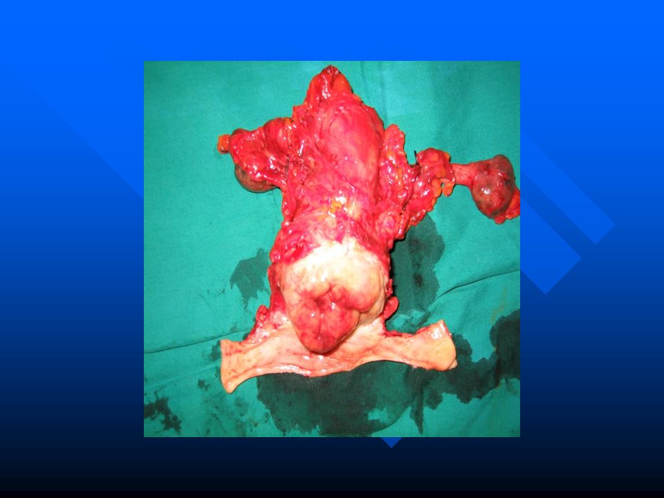
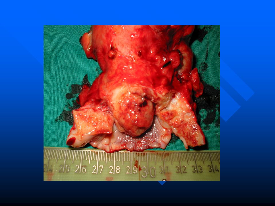
Kanker Serviks 1/Sellors/Ch. 8/p. 4/ Figures 3.5 and 8.8 Photos courtesy of Dr. J. Monsonego. 1. Reprinted with permission from Sellors JW, Sankaranarayanan R, eds. Colposcopy and Treatment of Cervical Intraepithelial Neoplasia. A Beginner’s Manual. Lyon, France: International Agency for Research on Cancer; 2003. 1/Sellors/Ch. 8/p. 2/¶ 5,6.

33
Stadium Kanker serviks
Stage I tumors: sel – sel kanker hanya terdapat dan terbatas pada leher rahim. Stage II tumors: sel tumor sudah menyebar di luar bagian dari leher rahim, sepert bagian atas dari vagina atau jaringan di sekitar leher rahim. Stage III tumors: tumor sudah menyebar di organ sekitar seperti bagian bawah dinding vagina, kelenjar getah bening terdekat atau jaringan lemak di sekitar leher rahim sampai mencapai dinding pelvis . Stage IV tumors: tumor menyebar ke organ – organ di sekitar organ genitalia ( yaitu kandung kemih atau usus) atau ke organ – organ di luar rongga pelvis . Meliputi penyebaran ke paru, hati atau tulang.
 atau ke organ – organ di luar rongga pelvis . Meliputi penyebaran ke paru, hati atau tulang.")
34
Stadium Klinis Kanker Leher Rahim
Stage I is carcinoma strictly confined to the cervix; extension to the uterine corpus should be disregarded. Stage IA: Invasive cancer identified only microscopically. All gross lesions even with superficial invasion are stage Ib cancers. Invasion is limited to measured stromal invasion with a maximum depth of 5 mm* and no wider than 7 mm. [Note: *The depth of invasion should not be more than 5 mm taken from the base of the epithelium, either surface or glandular, from which it originates. Vascular space involvement, either venous or lymphatic, should not alter the staging.] Stage IA1: Measured invasion of the stroma no greater than 3 mm in depth and no wider than 7 mm diameter. Stage IA2: Measured invasion of stroma greater than 3 mm but no greater than 5 mm in depth and no wider than 7 mm in diameter. Stage IB: Clinical lesions confined to the cervix or preclinical lesions greater than stage IA. Stage IB1: Clinical lesions no greater than 4 cm in size. Stage IB2: Clinical lesions greater than 4 cm in size. Stage II is carcinoma that extends beyond the cervix but has not extended onto the pelvic wall. The carcinoma involves the vagina, but not as far as the lower third. Stage IIA: No obvious parametrial involvement. Involvement of up to the upper two thirds of the vagina. Stage IIB: Obvious parametrial involvement, but not onto the pelvic sidewall. Stage III is carcinoma that has extended onto the pelvic sidewall. On rectal examination, there is no cancer-free space between the tumor and the pelvic sidewall. The tumor involves the lower third of the vagina. All cases with a hydronephrosis or nonfunctioning kidney should be included, unless they are known to be due to other causes. Stage IIIA: No extension onto the pelvic sidewall but involvement of the lower third of the vagina. Stage IIIB: Extension onto the pelvic sidewall or hydronephrosis or nonfunctioning kidney. Stage IV is carcinoma that has extended beyond the true pelvis or has clinically involved the mucosa of the bladder and/or rectum. Stage IVA: Spread of the tumor onto adjacent pelvic organs. Stage IVB: Spread to distant organs. Source: “FIGO Annual Report on The Results of Treatment in Gynaecological Cancer” Journal of Epidemiology and Biostatistics, (2001) vol. 6 no. 1, page 14.
 vol. 6 no. 1, page 14.")
35
Pengobatan Kanker Serviks
Surgery Radiation Therapy External Radiation Chemotherapy

36
Surgery Radikal Histerektomi, disertai
Pelvic lymph node diseksi (sentinal node biopsy) Para-aortic lymph node sampling
 Para-aortic lymph node sampling.")
39
Systemic Therapy and Radiation
Cisplatin Brachytherapy Pelvic Radioterapi

40
Terima Kasih

Presentasi serupa


>")


>")


