Upload presentasi
Presentasi sedang didownload. Silahkan tunggu

1
REGULASI SISTEM GASTROINTESTINAL
dr.Nuraiza Meutia, M.Biomed Dept. Fisiologi FK USU

2
The physiology of digestion
The gastrointestinal (GI) tract is the site of nutrient digestion and later absorption. Stretches from mouth to anus The GI tract moves and mixes food (motility) while also secreting chemicals to breakdown food. GI tract also eliminates waste.
 tract is the site of nutrient digestion and later absorption. Stretches from mouth to anus. The GI tract moves and mixes food (motility) while also secreting chemicals to breakdown food. GI tract also eliminates waste.")
3
Digestive System: Overview
Alimentary canal : mulut, pharynx, esophagus, lambung, usus halus dan usus besar. Accessory digestive organs : gigi geligi, lidah, kantung empedu, kelenjar liur, hati, dan pankreas

4
Digestive Process 6 aktivitas dalam proses digestif :
memasukkan, memindahkan, dan pencernaan secara mekanis, + pencernaan secara kimia, absorpsi, dan defekasi

5
Gastrointestinal Tract Activities
Ingestion (memasukkan) : taking food into the digestive tract Propulsion (memindahkan) : swallowing and peristalsis Mechanical digestion (pencernaan secara mekanis) : mengunyah, mencampur, dan mengaduk makanan.
 : taking food into the digestive tract. Propulsion (memindahkan) : swallowing and peristalsis. Mechanical digestion (pencernaan secara mekanis) : mengunyah, mencampur, dan mengaduk makanan.")
6
Gastrointestinal Tract Activities
Chemical digestion (pencernaan secara kimia) : catabolic breakdown of food Absorption : movement of nutrients from the GI tract to the blood or lymph Defecation : elimination of indigestible solid wastes
 : catabolic breakdown of food. Absorption : movement of nutrients from the GI tract to the blood or lymph. Defecation : elimination of indigestible solid wastes.")
7
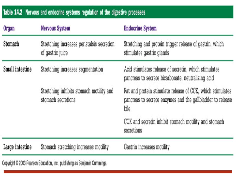
Pengaturan Kerja Saluran Cerna
3 hal yang mengatur kerja sistem pencernaan : 1. Persarafan Saraf otonom (ekstrinsik) dan enterik (intrinsik) 2. Hormonal 3. Mekanisme lokal Local messenger yang berespon thd perubahan pH atau stimulus kimia dan fisik.
 dan. enterik (intrinsik) 2. Hormonal. 3. Mekanisme lokal. Local messenger yang berespon thd perubahan pH atau stimulus kimia dan fisik.")
8
Pengaturan Kerja Saluran Cerna

9
Nervous Control of the GI Tract
Intrinsic controls Pleksus submukosa (plexus of Meissner) dan pleksus mienterik (plexus of Auerbach) Persarafan enterik lokal ini (local enteric plexuses / gut brain) menghasilkan Short reflexes
 dan pleksus mienterik (plexus of Auerbach) Persarafan enterik lokal ini (local enteric plexuses / gut brain) menghasilkan Short reflexes.")
10
Nervous Control of the GI Tract
Extrinsic controls Bagian dari simpatis dan parasimpatis yang besinap dengan neuron di dalam pleksus. Menghasilkan Long reflexes, within or outside the GI tract Juga melibatkan CNS.

11
Histology of the Alimentary Canal
Figure 23.6

12
Receptors of the GI Tract
Mechano- and chemoreceptors respond to: Stretch, osmolarity, and pH Presence of substrate, and end products of digestion They initiate reflexes that: Activate or inhibit digestive glands Mix lumen contents and move them along

13
Nervous Control of the GI Tract
Figure 23.4

14
CONTROL OF SALIVARY SECRETION
cerebral cortex other inputs conditioned reflex salivary centre in medulla simple reflex autonomic nerves pressure receptors and chemoreceptors in the mouth salivary glands salivary secretion

16
Persarafan pada setiap bagian GIT :
Mulut : Aktivitas Persarafan Keterangan Pergerakan rahang untuk mengunyah N. Trigeminus cabang mandibular (V3) - Pergerakan lidah mencampur N.Hipoglosus Sekresi saliva Simpatis (sup.cervical ganglion) dan Parasimpatis (N.VII,IX) Reseptor : N.V;N.VII,IX,X
 - Pergerakan lidah mencampur. N.Hipoglosus. Sekresi saliva. Simpatis (sup.cervical ganglion) dan Parasimpatis (N.VII,IX) Reseptor : N.V;N.VII,IX,X.")
17
Persarafan pada setiap bagian GIT :
Esofagus : Aktivitas Persarafan Menelan pada tahap oral Receptors : fauces, tonsils, soft palate, base of the tongue, post. pharyngeal wall. Afferent : N.VII, N.IX, N.X. Efferent : N.V, N.IX, N.X & N.IX. Menelan pada tahap faringeal Menelan pada tahap esofageal

18
Persarafan pada setiap bagian GIT :
Lambung s/d rectum : Aktivitas pada Parasimpatis Simpatis Lambung N. Vagus N. Splanchnic thoracic Usus halus Usus besar Rectum-Anus N. Splanchnic Pelvic N. Splanchnic Lumbar N.Pudendal (aktivitas motorik volunter)
")
19
The Physiology of digestion
GI tract propulsion: peristalsis Caused by circular and lengthwise muscle contraction Occurs in esophagus, stomach, small intestine Colon has occasional mass movements

20
Figure 14.3A

21
Kontraksi usus besar Mass movements Peristaltik haustra

22
The physiology of digestion
The mouth and stomach are connected by a tube called the esophagus. Epiglottis is a flap of tissue at the top of esophagus which prevents food from entering the trachea (windpipe).
.")
23
The physiology of digestion
GI tract control valves: sphincters Located throughout the intestinal tract Respond to inputs from nerves, hormones, hormone-like compounds and pressure around them Upper and lower esophageal sphincters

24
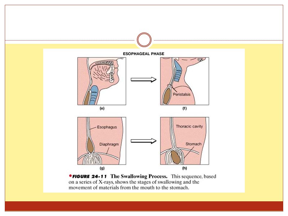
5 % bagian atas – otot rangka ½ bagian bawah – otot polos
Anatomi: Esofagus dimulai dari tingkat kartilago cricoid (C6) sampai ke bagian kardia lambung, sepanjang ± 25 cm pada pria dan 23 cm pada wanita. Diameter 2 cm. 5 % bagian atas – otot rangka ½ bagian bawah – otot polos di antaranya : campuran otot rangka & otot polos
 sampai ke bagian kardia lambung, sepanjang ± 25 cm pada pria dan 23 cm pada wanita. Diameter 2 cm. 5 % bagian atas – otot rangka. ½ bagian bawah – otot polos. di antaranya : campuran otot rangka & otot polos.")
25
Aktivitas Neuromuskular :
Selama fase esofageal, terbentuk “gelombang peristaltik primer” berkekuatan 100 cm tekanan H2O , yang menggerakkan bolus di sepanjang esofagus. Kecepatan 2-4 cm/dtk. 2. “Gelombang Sekunder" dapat terjadi bila terdapat peninggian tekanan di pertengahan esofagus- akibat adanya sisa makanan tertinggal di esofagus setelah selesainya berlangsung “gelombang primer”

26
3. “Gelombang Tertier" dapat timbul pada lansia dan keadaan patologis tertentu. Gelombang ini terjadi di bgn distal esofagus. 4. UES terbuka bila ada tekanan 25 cm tekanan air (cm H2O)dari bagian atas, tapi diperlukan lebih dari 100 cm tekanan air untuk membukanya dari bagian bawah. LES (sfinkter esofagus bawah) terbuka oleh 5-7 cm tekanan air dari bagian atas, tapi diperlukan lebih dari 25 cm tekanan air untuk menyebabkan refluks.
dari bagian atas, tapi diperlukan lebih dari 100 cm tekanan air untuk membukanya dari bagian bawah. LES (sfinkter esofagus bawah) terbuka oleh 5-7 cm tekanan air dari bagian atas, tapi diperlukan lebih dari 25 cm tekanan air untuk menyebabkan refluks.")
28
The anatomy and physiology of digestion
GI tract control valves: sphincters Pyloric sphincter Base of the stomach Allows stomach contents to enter intestine a few milliliters (1 tsp) at a time Ileocecal sphincter End of small intestine Prevents contents of large intestine from re-entering small intestine Two anal sphincters
 at a time. Ileocecal sphincter. End of small intestine. Prevents contents of large intestine from re-entering small intestine. Two anal sphincters.")
29
Stomach Four-cup holding tank Secretes acid and enzymes
Only proteins are significantly digested in the stomach Churns and mixes food Holds food for two to four hours Releases food “a little bit at a time” to the small intestine. Solid take longer than liquids and fat meal takes longer that CHO or protein

30
A closer look at the digestive process
Key digestive processes in the stomach Hormone-producing cells are released in response to thinking about or chewing food Stomach cells produce acid and enzymes Mucus lining protects stomach from acid Chyme is formed

31
Stomach Nerve supply – sympathetic and parasympathetic fibers of the autonomic nervous system

32
Kelenjar-kelenjar pada bagian Fundus dan Korpus Lambung
Mucous neck cells : acid mucus Parietal cells : HCl dan intrinsic factor Chief cells : pepsinogen Enteroendocrine cells : gastrin, histamine, endorphins, serotonin, cholecystokinin (CCK), dan somatostatin ke lamina propria
, dan somatostatin ke lamina propria.")
33
Response of the Stomach to Filling
Stomach pressure remains constant until about 1L of food is ingested Relative unchanging pressure results from reflex-mediated relaxation and plasticity Reflex-mediated events include: Receptive relaxation – as food travels in the esophagus, stomach muscles relax Adaptive relaxation – the stomach dilates in response to gastric filling Plasticity – intrinsic ability of smooth muscle to exhibit the stress-relaxation response

34
Gastric Contractile Activity
Peristaltic waves move toward the pylorus at the rate of 3 per minute This basic electrical rhythm (BER) is initiated by pacemaker cells (cells of Cajal) Most vigorous peristalsis and mixing occurs near the pylorus Chyme is either: Delivered in small amounts to the duodenum or Forced backward into the stomach for further mixing
 is initiated by pacemaker cells (cells of Cajal) Most vigorous peristalsis and mixing occurs near the pylorus. Chyme is either: Delivered in small amounts to the duodenum or. Forced backward into the stomach for further mixing.")
35
Gastric Contractile Activity
Figure 23.18

36
Regulation of Gastric Secretion
Pengeluaran cairan lambung diatur oleh mekanisme neural dan hormonal . Proses stimulasi atau inhibisi berlangsung dalam 3 fase : Cephalic (reflex) phase: sebelum makanan masuk Gastric phase: ketika makanan masuk ke lambung Intestinal phase: ketika sebagian dari makanan yang sudah dicerna masuk ke duodenum
 phase: sebelum makanan masuk. Gastric phase: ketika makanan masuk ke lambung. Intestinal phase: ketika sebagian dari makanan yang sudah dicerna masuk ke duodenum.")
37
1.Cephalic Phase Hal yang dapat menstimulasi :
Melihat atau memikirkan makanan Stimulasi reseptor pengecap atau penghidu Hal yang dapat menghambat : Depresi atau hilang nafsu makan Penurunan stimulasi parasimpatetik

38
Figure 24.15a

39
2.Gastric Phase Hal yang dapat menstimulasi : Stomach distension
Activation of stretch receptors (neural activation) Activation of chemoreceptors by peptides, caffeine, and rising pH Release of gastrin to the blood Hal yang dapat menghambat : A pH lower than 2 Emotional upset that overrides the parasympathetic division
 Activation of chemoreceptors by peptides, caffeine, and rising pH. Release of gastrin to the blood. Hal yang dapat menghambat : A pH lower than 2. Emotional upset that overrides the parasympathetic division.")
40
Figure 24.15b

41
3. Intestinal Phase Excitatory phase : low pH(keasaman meningkat); makanan yg sebagian telah dicerna memasuki duodenum dan mendorong /mengaktifkan kelenjar di lambung. Inhibitory phase : distensi duodenum, adanya lipid, acidic, atau kimus hipertonik, dan bahan iritan di duodenum (Initiates inhibition of local reflexes and vagal nuclei) (Closes the pyloric sphincter) (Releases enterogastrones that inhibit gastric secretion
; makanan yg sebagian telah dicerna memasuki duodenum dan mendorong /mengaktifkan kelenjar di lambung. Inhibitory phase : distensi duodenum, adanya lipid, acidic, atau kimus hipertonik, dan bahan iritan di duodenum. (Initiates inhibition of local reflexes and vagal nuclei) (Closes the pyloric sphincter) (Releases enterogastrones that inhibit gastric secretion.")
42
Figure 24.15c

43
Regulation of Gastric Emptying
Gastric emptying is regulated by: The neural enterogastric reflex Hormonal (enterogastrone) mechanisms These mechanisms inhibit gastric secretion and duodenal filling Carbohydrate-rich chyme quickly moves through the duodenum Fat-laden chyme is digested more slowly causing food to remain in the stomach longer
 mechanisms. These mechanisms inhibit gastric secretion and duodenal filling. Carbohydrate-rich chyme quickly moves through the duodenum. Fat-laden chyme is digested more slowly causing food to remain in the stomach longer.")
44
Gastric Motility what slows gastric emptying:
Hypertonic chyme duodenal pH <3.5 presence of amino acids and peptides in duodenum fatty acids and monoglycerides signals: what promotes emptying stomach distension and gastrin Acid in duodenum : decreases force of gastric contraction increases duodenal motility releases secretin which inhibits antral contraction and stimulates contraction of pyloric sphincter Fat products: partly from release of CCK from duodenum and jejunum and from GIP - decrease pyloric pump activity, slight increase in pyloric sphincter contraction - fats are emptied the slowest for gastric emptying, the volume of food in the stomach causes stretching of the gastric wall which elicits vagal and local myenteric reflexes that excite the pyloric pump. Gastrin - released with ingestion of meat - causes secretion of highly acidic gastric juice , and enhances the pyloric pump activity. Duodenal factors that inhibit emptying: enterogastric nervous reflexes - inhibitory reflexes from duodenal wall passing to stomach when volume of chyme in duodenum too much products of protein digestion - so that there will be enough time for protein digestion

45
Regulation of Gastric Emptying
Figure 23.19

46
Hunger contractions when stomach empty for long time rhythmic
cause tetanic contraction for 2-3 min most intense in young persons high degree of GI tonus low blood sugar level begins hrs after last food intake

47
Regulation and Mechanism of HCl Secretion
Sekresi HCl distimulasi oleh ACh, histamine, dan gastrin melalui sistem second-messenger Release of hydrochloric acid: Is low if only one ligand binds to parietal cells Is high if all three ligands bind to parietal cells Antihistamines block H2 receptors and decrease HCl release

48
Regulation and Mechanism of HCl Secretion

49
The physiology of digestion
Small intestine (about 10 feet) Duodenum, first 10 inches Jejunum, next four feet Ileum, last five feet Most digestion is completed in the jejunum Most digestive enzymes from intestine cells and pancreas Muscular contraction move and mix food Meal remains 3 to 10 hours with about 95% of the meal digested by the time it leaves
 Duodenum, first 10 inches. Jejunum, next four feet. Ileum, last five feet. Most digestion is completed in the jejunum. Most digestive enzymes from intestine cells and pancreas. Muscular contraction move and mix food. Meal remains 3 to 10 hours with about 95% of the meal digested by the time it leaves.")
50
A closer look at the digestive process
Key digestive processes in the small intestine Chyme triggers the release of bicarbonate Bicarbonate neutralizes acid Ulcer formation; intestine lacks mucus layer Pancreas releases enzymes Gallbladder releases bile

51
Small intestine: Site for most nutrient absorption
Key digestive processes in the small intestine Absorbs 95% of energy received from carbohydrate, protein, fat, and alcohol Large surface area promotes nutrient absorption Villi trap foodstuffs to enhance absorption Constant renewal of intestinal lining

52
Small intestine: Site for most nutrient absorption
Key digestive processes in the small intestine Various hormones and other substances participate in the digestive process Types and means of absorption Absorptive cells Absorption mechanisms Passive absorption Active absorption

53
Motility in the Small Intestine
The most common motion of the small intestine is segmentation It is initiated by intrinsic pacemaker cells (Cajal cells) Moves contents toward the ileocecal valve Local enteric neurons of the GI tract coordinate intestinal motility Cholinergic neurons cause: Contraction and shortening of the circular muscle layer Shortening of longitudinal muscle Distension of the intestine
 Moves contents toward the ileocecal valve. Local enteric neurons of the GI tract coordinate intestinal motility. Cholinergic neurons cause: Contraction and shortening of the circular muscle layer. Shortening of longitudinal muscle. Distension of the intestine.")
54
Q17: How are Proteins & Carbohydrates absorbed ?

55
Q18. How is fat Absorbed ?

56
The physiology of digestion
Large intestine: Colon 3 1/2 feet long Five sections: cecum, ascending, transverse, descending, and sigmoid Some digestion of leftover plant fibers Little other digestion Food remnants and wastes stay for about 24 to 72 hours

57
The large intestine completes absorption
5% of carbohydrate, protein, and fat escapes absorption Some water is present; absorbed in upper half of large intestine Sodium and potassium are absorbed in the upper half of large intestine Small amounts of undigested starches are absorbed Feces formed in last third of large intestine Insufficient enzyme production or incomplete digestion can cause abdominal discomfort

58
The physiology of digestion
Rectum and anus The Defecation Reflex Removes undigested faeces from the body. Stretch receptors in GIT wall detect distension of rectum. Parasympathetic reflex causes contractions of the sigmoid colon & rectum + relaxation of internal anal sphincter. External anal sphincter (under voluntary control) consciously relaxed if appropriate.
 consciously relaxed if appropriate.")
59
The Defecation Reflex Figure 24.25

60
TERIMA KASIH Refferences :
Elaine N.Marieb. Human Anatomy and Physiology. 5th Ed. Pearson Education Slide prepared by Vince Austin, University of Kentucky. Acknowledgement Figures have been taken from: Martini FH. Fundamentals of Anatomy and Physiology. 4th ed. Prentice Hall, 1998 Saladin KS. Anatomy and Physiology: The Unity of Form and Function. 2nd ed. McGraw-Hill, 2001

Presentasi serupa