Upload presentasi
Presentasi sedang didownload. Silahkan tunggu

1
RESPIRASI KUNCORO PUGUH S

2
RESPIRASI VENTILASI PARU :
Proses keluar / masuknya udara dari atm ke alveolus DIFUSI GAS : Difusi O2 &CO2 alv darah TRANSPOR GAS : Transpor O2 & CO2 alv darah PENGATURAN VENTILASI :

3
FUNGSI RESPIRASI UTAMA :
Membawa O2 sampai ke jaringan & mengambil CO2 dari jaringan SEKUNDER : Regulasi keasaman cairan ekstrasel Pengendalian suhu Eliminasi air Fonasi

4
The Respiratory System
The respiratory system works with the cardiovascular system to exchange gases between the air and blood (external respiration) and between blood and tissue fluids (internal respiration). Inspiration and expiration move air in and out of the lungs during breathing. Cellular respiration is the final destination where ATP is produced in cells.
 and between blood and tissue fluids (internal respiration). Inspiration and expiration move air in and out of the lungs during breathing. Cellular respiration is the final destination where ATP is produced in cells.")
5
Respiration Physiological process by which oxygen moves into an animal’s internal environment and carbon dioxide moves out Oxygen is needed for aerobic respiration Carbon dioxide is produced by same

6
Pressure Gradients Concentration gradients for gases
Gases diffuse down their pressure gradients Gases enter and leave the body by diffusing down pressure gradients across respiratory membranes

7
Atmospheric Pressure Pressure exerted by the weight of the air on objects on Earth’s surface At sea level = 760 mm Hg Oxygen is 21% of air; its partial pressure is about 160 mm Hg

8
Surface-to-Volume Ratio
As animal size increases, surface-to-volume ratio decreases Small, flat animals can use the body surface as their respiratory surface Larger animals have special structures to increase respiratory surface, such as gills or lungs

9
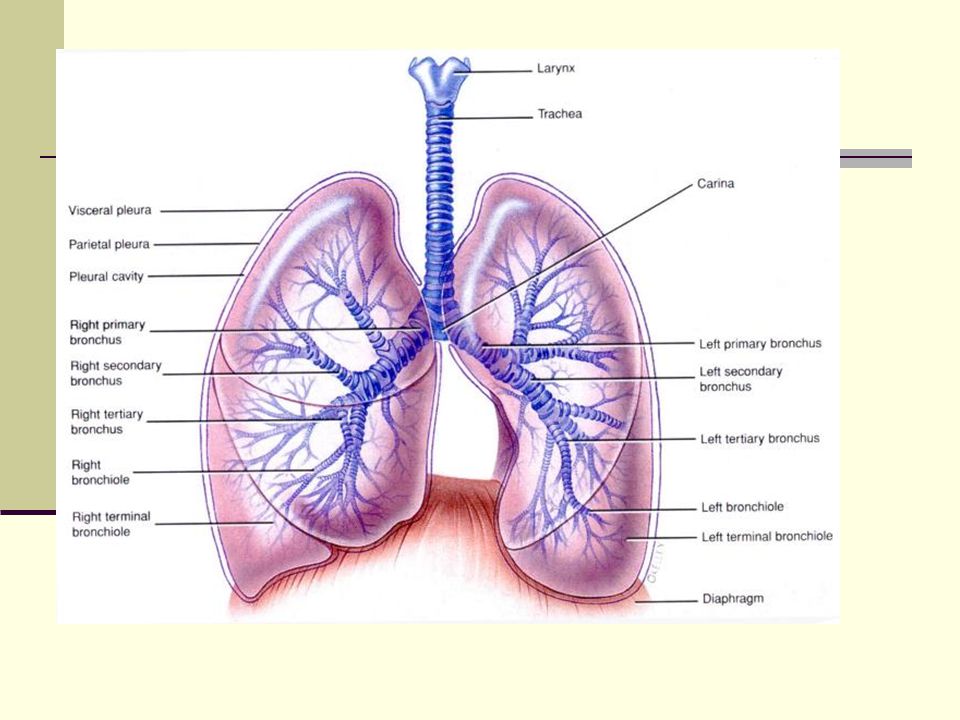
Ventilasi Paru Anatomi & Fisiologi Hidung Pharing Laring Trachea
Bronchus Bronchiolus Bronchiolus terminalis Bronchiolus Respiratorius Duktus alveolaris Saccus alveolaris Keterangan : Zona konduksi Zona transisional & Respirasi

11
HIDUNG Bulu rambut : Fungsi sbg filter ( 10 - 15µm) Sinus :
Membentuk ruang berkelok-kelok & pembuluh darah >> Fungsi untuk menjaga kelembaban dan suhu agar sesuai dengan tubuh

12
Sinusitis Pada Kuda yang menderita penyakit gigi terutama pada rahang bagian atas Pada Sapi yang mengalami dehorning (pemotongan tanduk)
 .")
13
Nostril = Cuping Hidung
Pada Kuda berfungsi untuk membedakan makanan Digunakan sebagai indikator hewan bila mengalami penyakit yang kronis, nostril bersisik dan kering

14
Pharing Pada Primata dan mamalia terdapat tiga saluran yaitu Saluran pernafasan, saluran makanan, saluran pendengaran

15
Nasal Cavity and Pharynx

16
Laring Tempat pembentukan suara

17
Speech Production Vocal cords stretch across laryngeal opening; opening between them is glottis Position of cords is varied to create different sounds

18
Vocal Folds

19
TRACHEA - BRONCHUS Tulang rawan berbentuk U dan Otot polos
Mucociliary escalator bergetar X/min sbg filter < 10 µm Bronchodilator : Adrenalin ( reseptor 2 ) P O2 P CO2 Bronchokonstriksi : Acethilcholine Histamine
 P O2. P CO2. Bronchokonstriksi : Acethilcholine. Histamine.")
20
Trachea Windpipe Divides to form Insert Fig 23.5 all but b
Primary bronchi Carina: Cough reflex Insert Fig 23.5 all but b

21
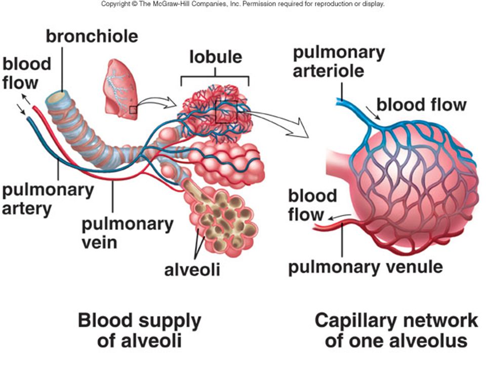
ALVEOLUS Mempunyai makrophag Mudah mengembang dan mengempis :
Jaringan elatik >> Surfaktan : fungsi untuk menurunkan tegangan permukaan paru, diproduksi sel alveolar tipe II

23
Bronchioles and Alveoli

25
Cutaway View of Alveolus
red blood cell air space inside alveolus (see next slide) pore for airflow between alveoli
 pore for airflow. between alveoli.")
26
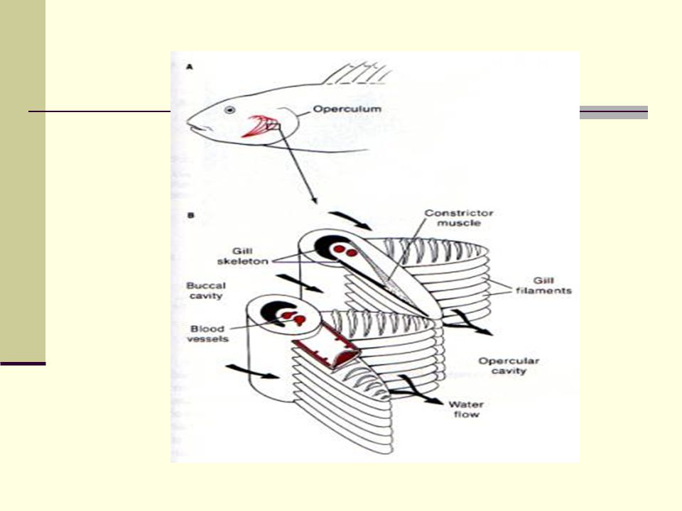
Fish Gills Most commonly internal
Water is drawn in through mouth and passed over gills water flows in through mouth FISH GILL water flows over gills, then out

27
Countercurrent Flow Blood flow runs in the opposite direction of water flow over the filaments This enhances movement of oxygen from water to blood respiratory surface direction of water flow direction of blood flow oxygenated blood back toward body oxygen-poor blood from deep in body

28
Vertebrate Lungs Originated in some fishes as outpouching from gut wall Allow gas exchange in oxygen-poor aquatic habitats and on land salamander reptile

29
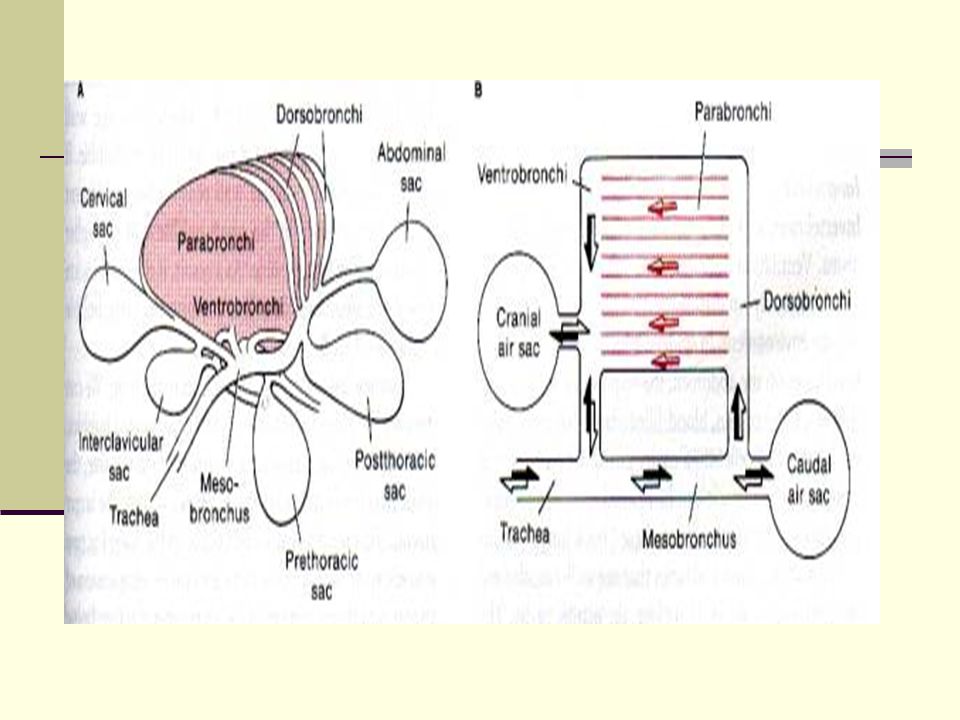
Avian Respiration Lungs are inelastic and connect to a series of air sacs Air is drawn continually though each lung air sacs air sacs lungs air sacs

32
Sistem sirkulasi Darah dari seluruh tubuh hewan akan melalui sistem sirkulasi kecil yaitu Atrium kanan -> Ventrikel kanan -> a. pulmonalis -> kapiler pulmonalis -> v. pulmonalis -> atrium kiri -> ventrikel kiri -> seluruh tubuh

34
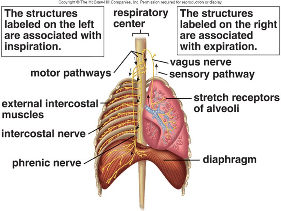
OTOT RESPIRASI INSPIRASI ( HISAP ) Diaphragma
M. Intercostalis eksterna M. Sternocleidomastoideus EKSPIRASI (HEMBUS ) M. Intercostalis interna Musculus abdominal
 M. Intercostalis interna. Musculus abdominal.")
35
There Inspiration and Expiration
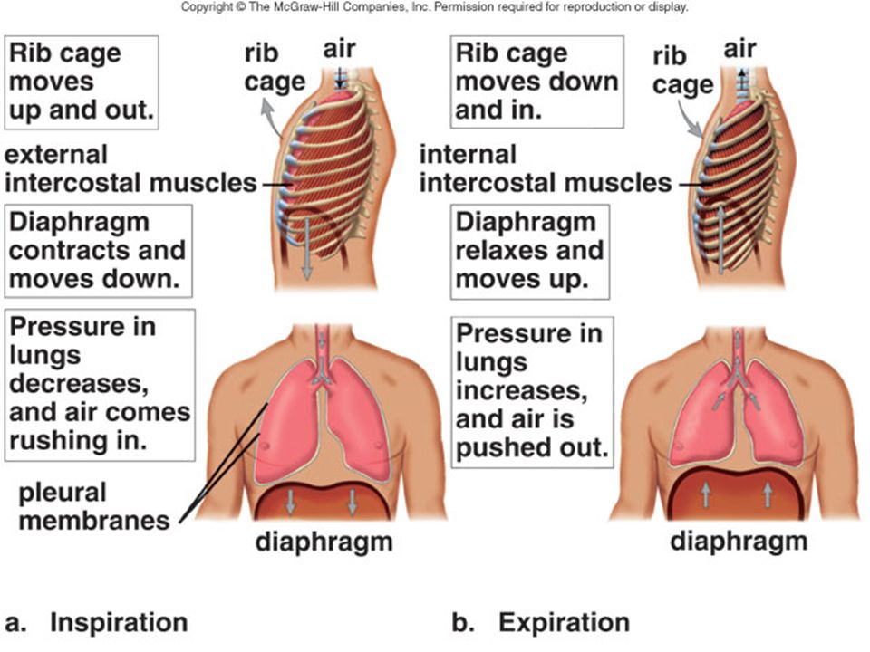
is a continuous column of air from the pharynx to the alveoli, and the lungs lie within the sealed-off thoracic cavity. The thoracic cavity is bounded by the rib cage and diaphragm. Pleural membranes line the thoracic cavity and lungs and the intrapleural pressure is lower than atmospheric pressure, keeping the lobules of the lungs from collapsing.

36
Tracheobronchial Tree

37
Inspiration When we inhale (inspiration) impulses from the respiratory center in the medulla oblongata cause the rib cage to rise and the diaphragm to lower, causing the thoracic cavity to expand. The negative pressure or partial vacuum in the alveoli causes the air to come in. Changing amounts of blood of CO2 and H+ increase breathing rate.
 impulses from the respiratory center in the medulla oblongata cause the rib cage to rise and the diaphragm to lower, causing the thoracic cavity to expand. The negative pressure or partial vacuum in the alveoli causes the air to come in. Changing amounts of blood of CO2 and H+ increase breathing rate.")
38
Inhalation Diaphragm flattens External intercostal muscles contract
Volume of thoracic cavity increases Lungs expand Air flows down pressure gradient into lungs

39
Expiration When we exhale (expiration), lack of impulses from the respiratory center allow the rib cage to lower and diaphragm to resume dome shape. Expiration is passive, while inspiration is active. The elastic recoil of the lungs causes expiration. A deep breath causes alveoli to stretch; stretch receptors then inhibit the respiratory center.

40
Normal (Passive) Exhalation
Muscles of inhalation relax Thoracic cavity recoils Lung volume decreases Air flows down pressure gradient and out of lungs

41
Active Exhalation Muscles in the abdomen and the internal intercostal muscles contract Contraction decreases thoracic cavity volume more than passive exhalation A greater volume of air must flow out to equalize intrapulmonary pressure with atmospheric pressure

44
Alveolar Pressure Changes

45
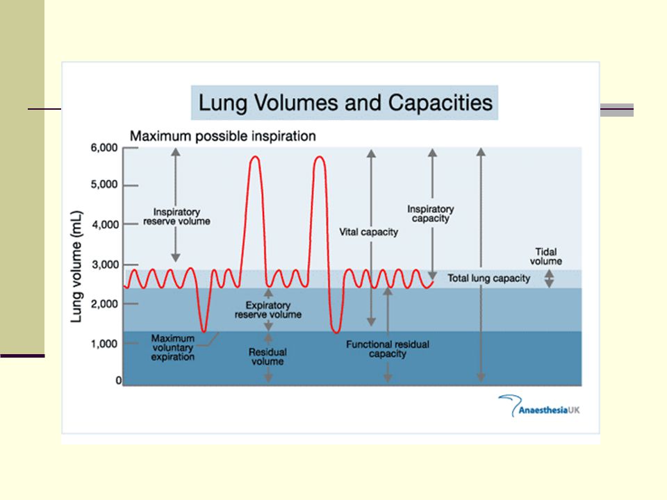
VOLUME & KAPASITAS PARU
VOLUME PARU Jumlah udara yang menempati petak-petak saluran pernafasan Contoh : Tidal Volume Inspiratory Reserve Volume Expiratory Reserve Volume Residual Volume

46
Kapasitas paru Penjumlahan dua atau lebih dari volume paru Contoh : Inspiratory Capacity Vital Capacity Functional Residual Capacity Total Lung Capacity Alat yang digunakan untuk mengukur SPIROMETER

48
Average Tidal Volume For Several Domestic Animal Species
BW ( Kg ) Condition Respiratory frequency ( breaths/min) Tidal Volume (ml/kg) Cattle Holstein cows Jersey cows Clinical index Horse Thoroughbred Clinical Index Dog Cat 516 405 - 486 19 12.6 13.8 3.7 Standing Anesthetized Resting Pentobarbital anesthesia 26 27 10 – 15 10 13.6 21.0 15.1 30 8.20 8.44 15.4 10.7 11.4 15.7 15 – 20 9.2
 Condition. Respiratory frequency. ( breaths/min) Tidal Volume. (ml/kg) Cattle. Holstein cows. Jersey cows. Clinical index. Horse. Thoroughbred. Clinical Index. Dog. Cat Standing. Anesthetized. Resting. Pentobarbital anesthesia – –")
49
Respiratory frequency for several animal species under different conditions
Cycles/minute Range Mean Horse Dairy cow Dairy Calf Swine Dog Cat Sheep Standing ( at rest ) Sternal recumbency Standing ( 52 Kg BW, 3 weeks age ) Lying down ( 52 Kg BW, 3 weeks age ) Lying down ( Kg BW) Sleeping ( at rest ) Sleeping Lying down, awake Standing, ruminating 10 – 14 26 – 35 24 – 50 18 – 22 21 – 25 32 – 58 18 – 25 20 – 34 16 – 25 20 – 40 12 29 35 20 22 40 21 24 31 25
 Sternal recumbency. Standing ( 52 Kg BW, 3 weeks age ) Lying down ( 52 Kg BW, 3 weeks age ) Lying down ( Kg BW) Sleeping ( at rest ) Sleeping. Lying down, awake. Standing, ruminating. 10 – – – – – – – – – –")
50
COMPLINCE ( DAYA KEMBANG)
PADA SISTEM PERNAFASAN TERDPAT TIGA MACAM COMPLIANCE : 1. COMPLIANCE PARU SAJA, YANG MERUPAKAN COMPLIANCE PARU BILA PARU DILEPAS DARI TORAKBESARNYA = 0,22 LITER / CM AIR 2. COMPLIANCE TORAK SAJA, YANG MERUPAKAN COMPLIANCE TORAK SAJA TANPA PARU 3. COMPLIANCE PARU-TORAK YANG MERUPAKAN COMPLIANCE SISTEM PERNAFASAN BESARNYA = 0,13 LITER /CM AIR

51
DEAD SPACE DEAD SPACE ANATOMIS
Ruang yang secara anatomis tidak ada pertukaran gas DEAD SPACE FISIOLOGIS Ruang yang secara anatomis tidak ada pertukaran gas ditambah alveoli yang tidak berfungsi karena difusi – atau pembululuh kapiler -

52
MINUTE VOLUME Volume udara yang terdapat dalam saluran pernafasan permenit pada pernafasan biasa (TV) TV X Frekuensi pernafasan ex : 500 ml X 12 /min = 6000ml/min

53
ALVEOLAR VENTILATION (TV - DEAD SPACE ) X FREKUENSI PERNAFASAN / MIN
Ex : ( 500 ml ml ) X 12 / min = 4200 ml/min
 X 12 / min. = 4200 ml/min.")
54
DIFUSI GAS Dipengaruhi oleh :
Tebal membran : semakin tebal akan semakin sulit Luas permukaan membran : semakin luas semakin cepat Koefisien difusi : kecepatan difusi melalui daerah tertentu pd jarak & perbedaan tertentu ex. O2=1,0 ;CO2=20,3 ;CO=0,81 ;N2=0,53 ;He=0,95 Gradien tekanan :bergerak dari tek. tinggi ke tek. rendah

55
Respiratory Membrane Area between an alveolus and a pulmonary capillary Oxygen and carbon dioxide diffuse across easily alveolar epithelium capillary endothelium fused basement membranes of both epithelial tissues

56
Changes in Partial Pressures

57
Skema difusi gas

58
TRANSPOR GAS Transpor O2 97% Oxyhemoglobin ( HbO2 ) 3% Plasma
Transpor CO2 67% Bicarbonate 24% Carbaminohemoglobin 9% Plasma

59
Oxygen Transport Most oxygen is carried bound to hemoglobin in red blood cells Hemoglobin has a great affinity for oxygen when it is at high partial pressure (in pulmonary capillaries) Lower affinity for oxygen in tissues, where partial pressure is low
 Lower affinity for oxygen in tissues, where partial pressure is low.")
60
Hemoglobin

61
Hemoglobin

62
Bicarbonate Formation
CO2 + H2O H2CO3 carbonic acid HCO3– bicarbonate + H+ Most carbon dioxide is transported as bicarbonate Some binds to hemoglobin Small amount dissolves in blood

63
TRANSPORT O2 DAN CO2 DARI JARINGAN KE SEL DRH

64
Carbon Dioxide Transport and Chloride Movement

65
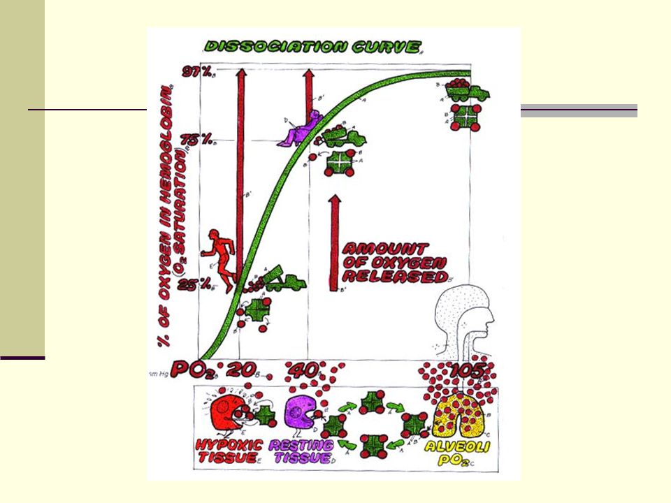
KURVA DISSOSIASI OXYHEMOGLOBINE
Hubungan antara PO2 plasma dengan % Saturasi Hb O2 bound to Hb % Saturasi Hb = X 100% O2 Capacity of Hb

67
KURVA DISOSIASI OKSIGEN
. Kurva ini dipengaruhi oleh suhu, PCO2, pH dan enzim DPG. Pengaruh mereka dapat dilihat pada kurva berikut : bergeser ke kanan bila : PCO2 meningkat pH turun Suhu naik DPG naik bergeser ke kiri PCO2 turun pH naik Suhu turun DPG turun PaO2 KIRI NORMAL KANAN

68
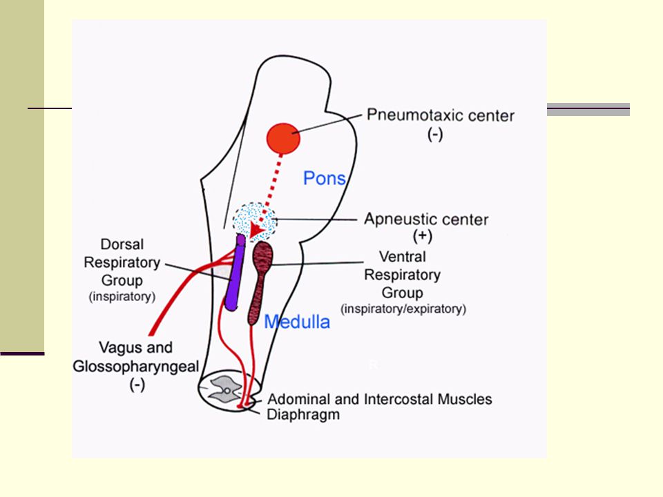
REGULASI PERNAFASAN Pusat pernafasan
Diatur oleh sekelompok neuron yang terletak dalam substasia retikularis dari medulla oblongata dan pons

70
Respiratory Structures in Brainstem

71
CHEMORESEPTOR ARTERIAL : Pada Arcus Aorta & A. Carotis CO2 meningkat
H2 menurun O2 menurun Central : di otak Karena CO2 dpt melewati sawar otak (Blood Brain Barrier System)
")
72
Control of Breathing Nervous system controls rhythm and magnitude of breathing Breathing is adjusted as a result of changes in Carbon dioxide levels Oxygen levels H+ levels Apnea = interrupted breathing

73
Modifying Respiration

74
Beberapa Istilah Cyanosis : kebiruan pd kulit yg disebabkan oleh jumlah Hb yg terdeoksigenasi meningkat di dlm p. drh kulit ( terutama kapiler) Eupnea : pernapasan normal Tachipnea : pernapasan cepat Bradypnea : pernapasan lambat Hipoksia : kekurangan oksigen di jaringan Hipoksemia : kekurangan oksigen di dalam darah

75
Regulation of Blood pH and Gases

76
Bronchitis Irritation of the ciliated epithelium that lines the bronchiole walls Air pollutants, smoking, or allergies can be the cause Excess mucus causes coughing, can harbor bacteria Chronic bronchitis scars and constricts airways

77
Emphysema An irreversible breakdown in alveolar walls
Lungs become inelastic May be caused by a genetic defect Most often caused by smoking

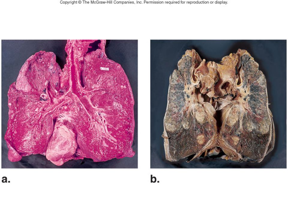
79
Fig

Presentasi serupa