Upload presentasi
Presentasi sedang didownload. Silahkan tunggu

1
SIKLUS SEL Made Pharmawati

2
Kontinuitas kehidupan
Didasarkan atas reproduksi sel atau pembelahan sel Figure 12.1

3
100 µm (a) Reproduction. An amoeba, a single-celled eukaryote, is dividing into two cells. Each new cell will be an individual organism (LM).
 Reproduction. An amoeba, a single-celled eukaryote, is dividing into two cells. Each new cell will be an individual organism (LM).")
4
Organisme multiseluler tergantung pada pembelahan sel untuk:
Perkembangan (dari sel yang terfertilisasi) Pertumbuhan Repair 20 µm 200 µm (b) Growth and development This micrograph shows a sand dollar embryo shortly after the fertilized egg divided, forming two cells (LM). (c) Tissue renewal. These dividing bone marrow cells (arrow) will give rise to new blood cells (LM). Figure 12.2 B, C
 Pertumbuhan. Repair. 20 µm. 200 µm. (b) Growth and development. This micrograph shows a sand dollar embryo shortly after the fertilized egg divided, forming two cells (LM). (c) Tissue renewal. These dividing bone marrow cells (arrow) will give rise to new blood cells (LM). Figure 12.2 B, C.")
5
Semua organisme kompleks berasal dari a single fertilized egg.
Melalui pembelahan sel, jumlah sel meningkat Sel kemudian terspesialisasi dan berubah menjadi fungsinya masing2

6
Tipe pembelahan sel Mitosis: Growth, development & repair
Asexual reproduction (yields identical cells) Occurs in somatic (body) cells Meiosis: Sexual reproduction (yields different cells) Occurs in specific reproductive cells So far, I’ve been talking about mitosis only What? Somatic (body) cells Why? Growth & development, repair lost or injured cells Allows many organisms to reproduce asexually But there is a 2nd type of cell division - meiosis that occurs only in select cells within certain tissue at particular phases of an organism’s lifetime. meiosis is involved with organisms that undergo SEXUAL REPRODUCTION --means of reducing number of chromosomes in sperm or egg so when combined through fertilization, the original number is restored -- reduction of genetic state from diploidy to haploidy necessary --occurs in reproductive organs in humans -- ovary & testis -- produces haploid cells called gametes (sperm, egg) --completed with fertilization of male gamete & female gamete to produce diploid zygote
 Occurs in somatic (body) cells. Meiosis: Sexual reproduction (yields different cells) Occurs in specific reproductive cells. So far, I’ve been talking about mitosis only. What Somatic (body) cells. Why Growth & development, repair lost or injured cells. Allows many organisms to reproduce asexually. But there is a 2nd type of cell division - meiosis that occurs only in select cells within certain tissue at particular phases of an organism’s lifetime. meiosis is involved with organisms that undergo SEXUAL REPRODUCTION. --means of reducing number of chromosomes in sperm or egg so when combined through fertilization, the original number is restored. -- reduction of genetic state from diploidy to haploidy necessary. --occurs in reproductive organs in humans -- ovary & testis -- produces haploid cells called gametes (sperm, egg) --completed with fertilization of male gamete & female gamete to produce diploid zygote.")
7
Pembelahan sel menghasilkan sel anak yang secara genetik identik
Phases of Mitosis 1. Prophase 2. Prometaphase 3. Metaphase 4. Anaphase 5. Telophase Pembelahan sel menghasilkan sel anak yang secara genetik identik Sel harus menduplikasikan material genetiknya Before they divide, ensuring that each daughter cell receives an exact copy of the genetic material, DNA

8
Distribution of Chromosomes During Cell Division
Pada persiapan cell division, DNA bereplikasi dan kromosom memadat Tiap chromosome yang terduplikasi memiliki dua sister chromatids, yang berpisah selama cell division Sentromer merupakan daerah ceking dari chromosome yang terduplikasi, diaman dua chromatids terikat dengan dekat

9
Figure 12.1 prophase 1X metahase Anaphase Telophase Cytokinesis

10
Cell division results in genetically identical daughter cells
Sel menduplikasikan material genetik sebelum membelah, utk memastikan bahwa tiap sel anak menerima copy DNA dengan tepat A cell’s endowment of DNA (its genetic information) is called its genome Molekul DNA dalam sel ter-pack menjadi kromosom
 is called its genome. Molekul DNA dalam sel ter-pack menjadi kromosom.")
11
Setiap spesies eukariot memiliki sejumlah tertentu kromosom pada nukleus
Sel somatik (nonreproductive) memiliki 2 set kromosom Gamet (reproductive cells: sperm and eggs) memiliki jumlah kromosom setengah jumlah kromosom sel somatik Kromosom eukaryotik terdiri dari kromatin, sebuah komplex DNA dan protein yang memampat selama pembelahan sel
 memiliki 2 set kromosom. Gamet (reproductive cells: sperm and eggs) memiliki jumlah kromosom setengah jumlah kromosom sel somatik. Kromosom eukaryotik terdiri dari kromatin, sebuah komplex DNA dan protein yang memampat selama pembelahan sel.")
12
Fig. 19.1, p. 345: cartoon (left), EM (right)
Chromatin = helix wrapped around protein -- giving bead-like structure These proteins are called ‘histones’ Nucleosome = DNA + histone complex Chromatin = a string of these beads (thread-like) Chromosome = looped and compacted chromatin
 Chromosome = looped and compacted chromatin.")
13
0.5 µm Chromosome duplication (including DNA synthesis) Centromere Separation of sister chromatids Sister chromatids Centromeres A eukaryotic cell has multiple chromosomes, one of which is represented here. Before duplication, each chromosome has a single DNA molecule. Once duplicated, a chromosome consists of two sister chromatids connected at the centromere. Each chromatid contains a copy of the DNA molecule. Mechanical processes separate the sister chromatids into two chromosomes and distribute them to two daughter cells. Figure 12.4

14
Pembelahan sel pada eukariot terdiri dari:
Mitosis, the division of the nucleus Cytokinesis, the division of the cytoplasm Gamet diproduksi dalam pembelahan sel yang disebut meiosis Meiosis menghasilkan sel anak yang tidak identik dengan induk yaitu hanya memiliki 1 set kromosom

15
Pada pembelahan sel, fase mitosis bergantian dengan interfase
Pada tahun 1882, ahli anatomi Jerman Walther Flemming mengembangkan pewarna untuk mengamati kromosom selama mitosis dan sitokinesis Bagi Flemming, terlihat sel tumbuh membesar Sekarang dapat diketahui banyak peristiwa kritis terjadi selama tahapan siklus sel

17
Fase-fase dalam siklus sel
Siklus sel terdiri dari Fase mitosis Interphase INTERPHASE G1 S (DNA synthesis) G2 Cytokinesis Mitosis MITOTIC (M) PHASE Figure 12.5 Interphase G1 phase S phase G2 phase The mitotic phase mitosis cytokinesis
 G2. Cytokinesis Mitosis. MITOTIC (M) PHASE. Figure Interphase. G1 phase. S phase. G2 phase. The mitotic phase. mitosis. cytokinesis.")
18
Phases of the Cell Cycle
Siklus sel terdiri dari Mitotic (M) phase (mitosis and cytokinesis) Interphase (cell growth and copying of chromosomes in preparation for cell division) Interphase (terdiri dari sekitar 90% dari siklus sel) yang dapat dibagi dalam sub fase: G1 phase (“first gap”) S phase (“synthesis”) G2 phase (“second gap”)
 phase (mitosis and cytokinesis) Interphase (cell growth and copying of chromosomes in preparation for cell division) Interphase (terdiri dari sekitar 90% dari siklus sel) yang dapat dibagi dalam sub fase: G1 phase ( first gap ) S phase ( synthesis ) G2 phase ( second gap )")
19
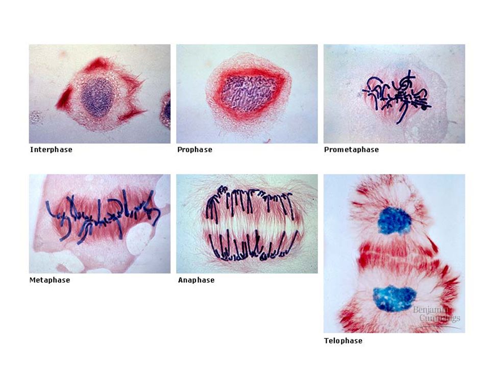
Mitosis terdiri dari 5 phases
Prophase Prometaphase G2 OF INTERPHASE PROPHASE PROMETAPHASE Centrosomes (with centriole pairs) Chromatin (duplicated) Early mitotic spindle Aster Centromere Fragments of nuclear envelope Kinetochore Nucleolus Nuclear envelope Plasma membrane Chromosome, consisting of two sister chromatids Kinetochore microtubule Figure 12.6 Nonkinetochore microtubules
 Chromatin (duplicated) Early mitotic spindle. Aster. Centromere. Fragments of nuclear envelope. Kinetochore. Nucleolus. Nuclear envelope. Plasma membrane. Chromosome, consisting of two sister chromatids. Kinetochore microtubule. Figure Nonkinetochore microtubules.")
20
Metaphase Anaphase Telophase Figure 12.6 Metaphase plate
Centrosome at one spindle pole Daughter chromosomes METAPHASE ANAPHASE TELOPHASE AND CYTOKINESIS Spindle Metaphase plate Nucleolus forming Cleavage furrow Nuclear envelope forming Figure 12.6

21
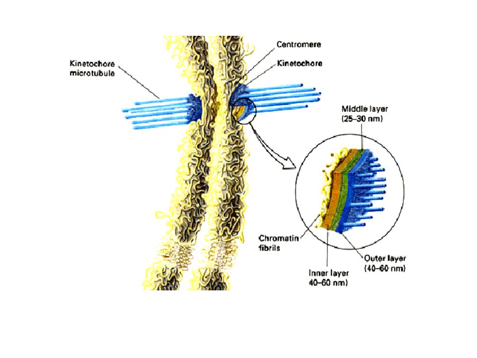
Spindle muncul dari sentromer
The mitotic spindle mikrotubul yang mengontrol pergerakan kromosom selama mitosis Spindle muncul dari sentromer spindle microtubules asters

22
Perakitan spindle microtubules dimulai dari sentrosom - microtubule organizing center
Sentrosom bereplikasi membentuk dua sentrosom yang bermigrasi ke kutub yang berlawanan, dan spindle microtubules tumbuh dari sentrosom Aster (a radial array of short microtubules) muncul dari tiap sentrosom
 muncul dari tiap sentrosom.")
23
The Spindle Spindle memiliki struktur seperti web terbuat dari microtubule . Sangat penting pada mitosis karena mengatur kromosom untuk berada pada posisi yang benar Mitotic center Click the return button to return to the prophase slide. Or the house button to return to the main menu. The purpose of the spindle is to organise the chromosomes during mitosis. It is a cradle of microtubule fibres which cause constriction around the centre of the cell, causing the cytoplasm to split. A cell at metaphase a spindle Microtubule

24
Some spindle microtubules
Berikatan dengan kinetochores chromosomes Centrosome Aster Sister chromatids Metaphase Plate Kinetochores Overlapping nonkinetochore microtubules Kinetochores microtubules Chromosomes Microtubules 0.5 µm 1 µm Figure 12.7

25
Chromosomes attached to spindle during nuclear division

26
Two kinds of microtubules
Kinetochore microtubules : berikatan dengan kinetochores chromosomes dan menggerakkan kromosom ke daerah metafase Nonkinetochores: overlap satu sama lain tetapi tidak berikatan dengan chromosome 1 µm

28
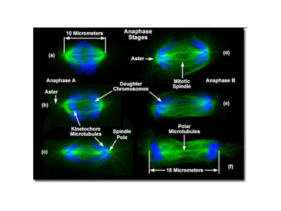
Pada anafase, sister chromatid berpisah
Dan bergerak sepanjang kinetochore microtubules menuju arah berlawanan ujung sel Spindle pole Kinetochore Figure 12.8

29
Initiation of Anaphase

30
The microtubules shorten by depolymerizing at their kinetochore ends
Nonkinetechore microtubules from opposite poles Overlap and push against each other, elongating the cell In telophase Genetically identical daughter nuclei form at opposite ends of the cell

32
Cytokinesis is the splitting of cytoplasm
Division of the cytoplasm Mitosis is the splitting of the nucleus. Cytokinesis is the splitting of cytoplasm It usually begins during ANAPHASE

33
Pada sel hewan Cytokinesis terjadi oleh proses yang disebut cleavage, membentuk sebuah a cleavage furrow Cleavage furrow Contractile ring of microfilaments Daughter cells 100 µm (a) Cleavage of an animal cell (SEM)
 Cleavage of an animal cell (SEM)")
34
Pada sel tumbuhan, selama cytokinesis
Terbentuk plat sel (cell plate) Daughter cells 1 µm Vesicles forming cell plate Wall of patent cell Cell plate New cell wall (b) Cell plate formation in a plant cell (SEM) Figure 12.9 B
 Daughter cells. 1 µm. Vesicles forming cell plate. Wall of patent cell. Cell plate. New cell wall. (b) Cell plate formation in a plant cell (SEM) Figure 12.9 B.")
35
Mitosis in a plant cell Nucleus Chromatine condensing Chromosome
1 Prophase. The chromatin is condensing. The nucleolus is beginning to disappear. Although not yet visible in the micrograph, the mitotic spindle is staring to from. Prometaphase. We now see discrete chromosomes; each consists of two identical sister chromatids. Later in prometaphase, the nuclear envelop will fragment. Metaphase. The spindle is complete, and the chromosomes, attached to microtubules at their kinetochores, are all at the metaphase plate. Anaphase. The chromatids of each chromosome have separated, and the daughter chromosomes are moving to the ends of cell as their kinetochore microtubles shorten. Telophase. Daughter nuclei are forming. Meanwhile, cytokinesis has started: The cell plate, which will divided the cytoplasm in two, is growing toward the perimeter of the parent cell. 2 3 4 5 Nucleus Nucleolus Chromosome Chromatine condensing Figure 12.10

36
Purpose of Interphase – to duplicate cell contents; 90% of the cell’s growth cycle
Purpose of Mitosis – to divide the genetic material into exact two halves Purpose of Cytokinesis – to divide all other contents (except nucleus) into two cells
 into two cells.")
37
Prokaryotes (bacteria and archaea) reproduce by a type of cell division called binary fission
In binary fission – The bacterial chromosome replicates – The two daughter chromosomes actively move apart

38
Chromosome replication begins. Soon thereafter,
LE 12-11_1 Cell wall Origin of replication Plasma membrane E. coli cell Bacterial chromosome Chromosome replication begins. Soon thereafter, one copy of the origin moves rapidly toward the other end of the cell. Two copies of origin

39
Chromosome replication begins. Soon thereafter,
LE 12-11_2 Cell wall Origin of replication Plasma membrane E. coli cell Bacterial chromosome Chromosome replication begins. Soon thereafter, one copy of the origin moves rapidly toward the other end of the cell. Two copies of origin Origin Origin Replication continues. One copy of the origin is now at each end of the cell.

40
Chromosome replication begins. Soon thereafter,
LE 12-11_3 Cell wall Origin of replication Plasma membrane E. coli cell Bacterial chromosome Chromosome replication begins. Soon thereafter, one copy of the origin moves rapidly toward the other end of the cell. Two copies of origin Origin Origin Replication continues. One copy of the origin is now at each end of the cell. Replication finishes. The plasma membrane grows inward, and new cell wall is deposited. Two daughter cells result.

41
A comparison of mitosis and meiosis

42
A comparison of mitosis and meiosis: summary

Presentasi serupa


>")


