Upload presentasi
Presentasi sedang didownload. Silahkan tunggu

1
Biochemistry Departement Medical Faculty Of Andalas University
BLOOD COMPOSITION Dr. Husnil Kadri, M.Kes Biochemistry Departement Medical Faculty Of Andalas University Padang

2
Fungsi Utama Darah 1. Respirasi; pengangkutan O2 dan CO2 2. Nutrisi;
pengangkutan hasil absorpsi usus 3. Ekskresi; pengangkutan sisa metabolik ke ginjal, paru-paru, kulit, & usus

3
Fungsi Utama Darah 4. Keseimbangan asam-basa 5. Keseimbangan air;
antara sirkulasi darah dan jaringan 6. Pengaturan suhu tubuh 7. Pertahanan terhadap infeksi; oleh sel darah putih & antibodi

4
Fungsi Utama Darah 8. Pengangkutan hormon & pengaturan metabolisme
9. Pengangkutan metabolit 10. Koagulasi

5
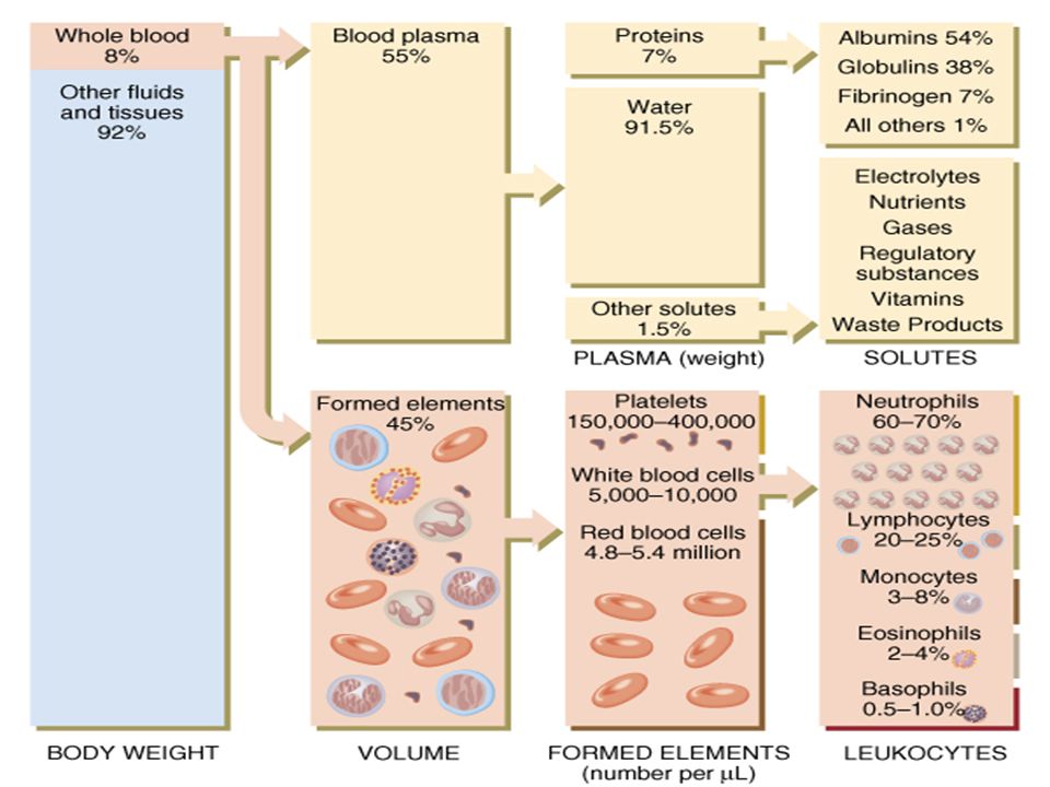
Components of Whole Blood
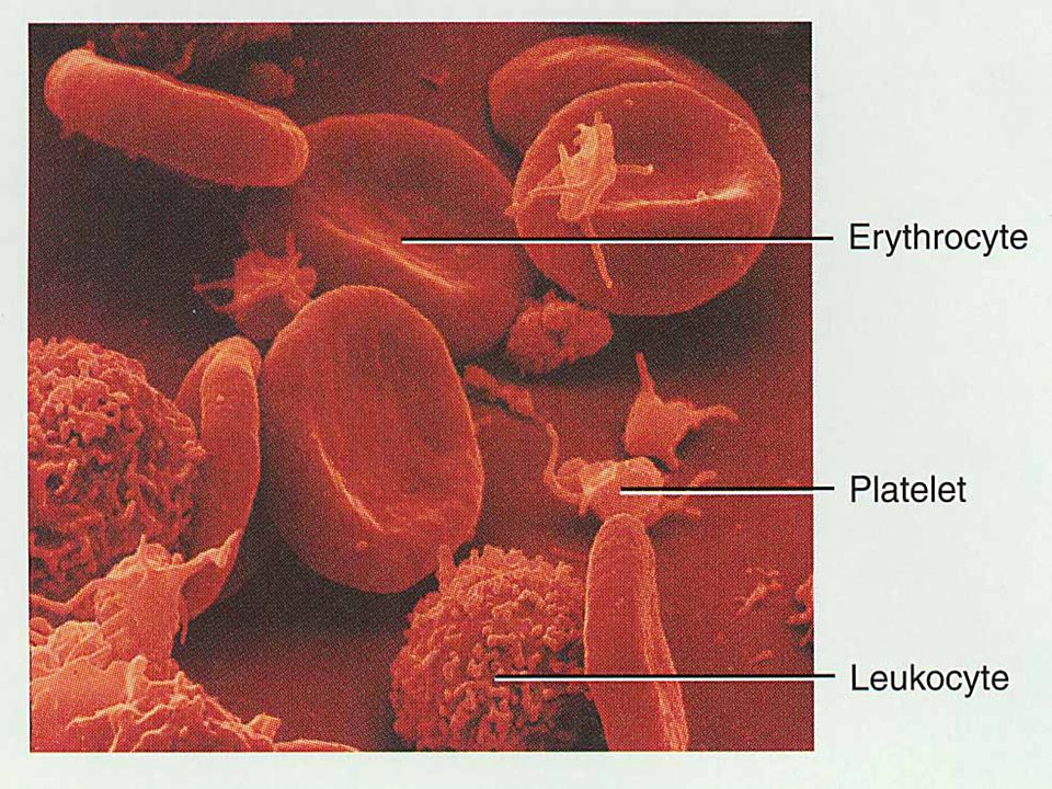
Plasma (55% of whole blood) Buffy coat: leukocyctes and platelets (<1% of whole blood) Formed elements Erythrocytes (45% of whole blood) 1 Withdraw blood and place in tube 2 Centrifuge Hematocrit Males: 47% ± 5% Females: 42% ± 5%
 Buffy coat: leukocyctes and platelets (<1% of whole blood) Formed elements. Erythrocytes (45% of whole blood) 1. Withdraw blood and place in tube. 2. Centrifuge. Hematocrit. Males: 47% ± 5% Females: 42% ± 5%")
8
Physical Characteristics of Blood
Average volume of blood: 5–6 L for males; 4–5 L for females (Normovolemia) Hypovolemia - low blood volume Hypervolemia - high blood volume Viscosity (thickness) (where water = 1) The pH of blood is 7.35–7.45; x = 7.4 Osmolarity = 300 mOsm or 0.3 Osm This value reflects the concentration of solutes in the plasma Salinity = 0.85% Reflects the concentration of NaCl in the blood Temperature is 38C, slightly higher than “normal” body temperature Blood accounts for approximately 8% of body weight
 Hypovolemia - low blood volume. Hypervolemia - high blood volume. Viscosity (thickness) (where water = 1) The pH of blood is 7.35–7.45; x = 7.4. Osmolarity = 300 mOsm or 0.3 Osm. This value reflects the concentration of solutes in the plasma. Salinity = 0.85% Reflects the concentration of NaCl in the blood. Temperature is 38C, slightly higher than normal body temperature. Blood accounts for approximately 8% of body weight.")
9
Components of Blood 55% plasma 45% cells 99% RBCs
< 1% WBCs and platelets

11
Blood Plasma Blood plasma components: Water = 90-92% Proteins = 6-8%
Albumins; maintain osmotic pressure of the blood Globulins Alpha and beta globulins are used for transport purposes Gamma globulins are the immunoglobulins (IgG, IgA, etc) Fibrinogen; a clotting protein Organic nutrients – glucose, carbohydrates, amino acids Electrolytes – sodium, potassium, calcium, chloride, bicarbonate Nonprotein nitrogenous substances – lactic acid, urea, creatinine Respiratory gases – oxygen and carbon dioxide
 Fibrinogen; a clotting protein. Organic nutrients – glucose, carbohydrates, amino acids. Electrolytes – sodium, potassium, calcium, chloride, bicarbonate. Nonprotein nitrogenous substances – lactic acid, urea, creatinine. Respiratory gases – oxygen and carbon dioxide.")
12
Formed Elements Formed elements comprise 45% of blood
Erythrocytes, leukocytes, and platelets make up the formed elements Only WBCs are complete cells RBCs have no nuclei or organelles, and platelets are just cell fragments Most formed elements survive in the bloodstream for only a few days Most blood cells do not divide but are renewed by cells in bone marrow

13
Erythrocytes (RBCs) Biconcave disc
Folding increases surface area (30% more surface area) Plasma membrane contains spectrin Give erythrocytes their flexibility Anucleate, no centrioles, no organelles End result - no cell division No mitochondria means they generate ATP anaerobically Prevents consumption of O2 being transported Filled with hemoglobin (Hb) - 97% of cell contents Hb functions in gas transport Hb + O HbO2 (oxyhemoglobin) Most numerous of the formed elements Females: 4.3–5.2 million cells/cubic millimeter Males: 5.2–5.8 million cells/cubic millimeter
 Plasma membrane contains spectrin. Give erythrocytes their flexibility. Anucleate, no centrioles, no organelles. End result - no cell division. No mitochondria means they generate ATP anaerobically. Prevents consumption of O2 being transported. Filled with hemoglobin (Hb) - 97% of cell contents. Hb functions in gas transport. Hb + O2 HbO2 (oxyhemoglobin) Most numerous of the formed elements. Females: 4.3–5.2 million cells/cubic millimeter. Males: 5.2–5.8 million cells/cubic millimeter.")
14
Erythrocytes (RBCs) Figure 17.3
 Figure 17.3")
15
Erythrocyte Function Erythrocytes are dedicated to respiratory gas transport Hemoglobin reversibly binds with oxygen and most oxygen in the blood is bound to hemoglobin Composition of hemoglobin A protein called globin made up of two alpha and two beta chains A heme molecule Each heme group bears an atom of iron, which can bind to one oxygen molecule Each hemoglobin molecule thus can transport four molecules of oxygen

16
Structure of Hemoglobin
Figure 17.4

17
Hemoglobin Satu mol. Hb dewasa (HbA) mempunyai; - 4 gugus heme
- Setiap heme mengandung 1 ion Fe2+ - 4 subunit protein globin - Setiap subunit mengikat 1 mol. O2 - 1 mol. Globin mengikat 1 mol. CO2 Subunit rantai terdiri dari 2 a dan 2 b; - a masing-masing = 141 asam amino - b masing-masing = 146 asam amino

18
Hemoglobin Oxyhemoglobin – hemoglobin bound to oxygen
Oxygen loading takes place in the lungs Deoxyhemoglobin – hemoglobin after oxygen diffuses into tissues (reduced Hb) Carbaminohemoglobin – hemoglobin bound to carbon dioxide Carbon dioxide loading takes place in the tissues
 Carbaminohemoglobin – hemoglobin bound to carbon dioxide. Carbon dioxide loading takes place in the tissues.")
19
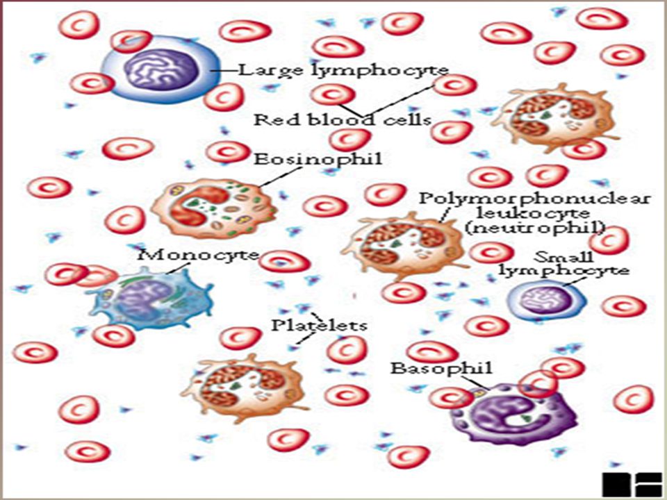
WBC Anatomy and Types All WBCs (leukocytes) have a nucleus and no hemoglobin Granular or agranular classification based on presence of cytoplasmic granules made visible by staining granulocytes are neutrophils, eosinophils or basophils agranulocytes are monocyes or lymphocytes

20
Differential WBC Count
Detection of changes in numbers of circulating WBCs (percentages of each type) indicates infection, poisoning, leukemia, chemotherapy, parasites or allergy reaction Normal WBC counts neutrophils 60-70% (up if bacterial infection) lymphocyte 20-25% (up if viral infection) monocytes % (up if fungal/viral infection) eosinophil % (up if parasite or allergy reaction) basophil <1% (up if allergy reaction or hypothyroid)
 indicates infection, poisoning, leukemia, chemotherapy, parasites or allergy reaction. Normal WBC counts. neutrophils 60-70% (up if bacterial infection) lymphocyte 20-25% (up if viral infection) monocytes % (up if fungal/viral infection) eosinophil % (up if parasite or allergy reaction) basophil <1% (up if allergy reaction or hypothyroid)")
21
Neutrophils (Granulocyte)
Polymorphonuclear Leukocytes or Polys Nuclei = 2 to 5 lobes connected by thin strands older cells have more lobes young cells called band cells because of horseshoe shaped nucleus (band) Fine, pale lilac practically invisible granules Diameter is microns 60 to 70% of circulating WBCs
 Fine, pale lilac practically invisible granules. Diameter is microns. 60 to 70% of circulating WBCs.")
22
Eosinophils (Granulocyte)
Nucleus with 2 or 3 lobes connected by a thin strand Large, uniform-sized granules stain orange-red with acidic dyes do not obscure the nucleus Diameter is 10 to 12 microns 2 to 4% of circulating WBCs

23
Basophils (Granulocyte)
Large, dark purple, variable-sized granules stain with basic dyes obscure the nucleus Irregular, s-shaped, bilobed nuclei Diameter is 8 to 10 microns Less than 1% of circulating WBCs

24
Lymphocyte (Agranulocyte)
Dark, oval to round nucleus Cytoplasm sky blue in color amount varies from rim of blue to normal amount Small cells microns in diameter Large cells microns in diameter increase in number during viral infections 20 to 25% of circulating WBCs

25
B cells responsible for production of antibodies
Lymphocytes B cells - responsible for humoral immunity T cells - responsible for cell mediated immunity B cells responsible for production of antibodies Receptor matches antigen Cells multiply Antibodies

26
T cells Cytotoxic T cells (Killer T cells) Helper T cells
Bind to cytotoxic cells (eg infected by virus) Swell Release toxins into cytoplasm Helper T cells Most numerous Activate B cells, killer T cells Stimulate macrophages Suppressor T cells Regulate activities of other cell types
 Swell. Release toxins into cytoplasm. Helper T cells. Most numerous. Activate B cells, killer T cells. Stimulate macrophages. Suppressor T cells. Regulate activities of other cell types.")
27
Monocyte (Agranulocyte)
Nucleus is kidney or horse-shoe shaped Largest WBC in circulating blood does not remain in blood long before migrating to the tissues differentiate into macrophages fixed group found in specific tissues alveolar macrophages in lungs kupffer cells in liver wandering group gathers at sites of infection Diameter is microns Cytoplasm is a foamy blue-gray 3 to 8% o circulating WBCs

28
Emigration & Phagocytosis in WBCs

29
UNSUR SELULAR DALAM RESPON IMUN
Jalur limfoid yang membentuk limfosit dan subsetnya Jalur mieloid yang membentuk sel-sel fagosit mononuklear & polimorfonuklear (PMN). PMN terdiri dari: neutrofil, eosinofil, basofil
. PMN terdiri dari: neutrofil, eosinofil, basofil.")
30
Platelets Platelets are fragments of mega-karyocytes
Their granules contain serotonin, Ca2+, enzymes, ADP, and platelet-derived growth factor (PDGF) Platelets function in the clotting mechanism by forming a temporary plug that helps seal breaks in blood vessels Platelets not involved in clotting are kept inactive by Nitric Oxide (NO) and prostaglandins
 Platelets function in the clotting mechanism by forming a temporary plug that helps seal breaks in blood vessels. Platelets not involved in clotting are kept inactive by Nitric Oxide (NO) and prostaglandins.")
31
Protein Plasma - Bagian utama unsur padat dalam plasma. - Konsentrasi total protein plasma + 7-7,5 g/dl. - Berbagai protein plasma dapat dipisahkan menurut karakteristik kelarutannya. - Metode pemisahan tsb antara lain; 1. Salting-out (Na2SO4 23%, dll) 2. Elektroforesis
 2. Elektroforesis.")
32
Zone Electrophoresis of Plasma Proteins
- + globulins albumin g b a1 a2 pI 6.0 5.6 5.1 4.7

33
Protein Plasma Sebagian besar disintesis di hepar. Umumnya disintesis sbg preprotein pada poliribosom terikat membran. Preprotein akan mengalami modifikasi pascatranslasi. Hampir semuanya berupa glikoprotein, kecuali albumin. Bersifat polimorfisme (ciri bawaan pd populasi dgn sedikitnya 2 macam fenotipe). contoh; gol. Darah ABO
. contoh; gol. Darah ABO.")
34
Plasma Proteins More than 200 Most abundant Albumin g/100 mL - g-globulins - ~1 g/100 mL fibrinogen g/100 mL

35
Albumin - Merupakan protein utama dalam plasma. - Mempertahankan 75-80% tekanan osmotik. - Berfungsi mengikat berbagai macam ligand, seperti; asam lemak bebas, Ca, Cu, Zn, hormon steroid, bilirubin, metheme

36
Albumin - Albumin juga dapat mengikat obat-an, seperti; sulfonamid, penisilin-G, dikumarol, aspirin - Penyakit hepar akan memperlihatkan rasio albumin/globulin yang menurun.

37
Transferin Adalah b1-globulin berbentuk glikoprotein yang disintesis di hepar. Berfungsi sebagai alat transpor besi (Fe3+) untuk dibawa ke jaringan. Jika besi tidak diikat oleh transferin, maka akan menjadi prooksidan.

38
Ceruloplasmin Protein ini adalah a2-globulin yang mengandung 90% Cu plasma. Tetapi 10% Cu terikat longgar pd albumin, sehingga mudah dilepas ke jaringan. Ceruloplasmin mengandung ferroksidase yang mengkatalisis ion Fe2+ --> Fe3+, karena hanya ion Fe3+ yang mampu berikatan dgn apotransferin.

39
g-Globulins 20% of plasma proteins “g” refers to electrophoretic mobility Represents a group of proteins of variable structure immunoglobulins Main functional task is immunochemical Antibodies - combine with specific antigens

40
Disintesis dalam sel plasma.
Imunoglobulin Plasma Disintesis dalam sel plasma. Sel plasma adalah turunan Sel-b yang mensintesis dan mensekresikan imuno- globulin sebagai respon terhadap pajanan berbagai antigen. Semua imunoglobulin mengandung paling kurang 2 rantai ringan dan 2 rantai berat.

41
Classes of Immunoglobulins
IgG – Identifies microorganisms for engulfment or lysis IgE – Inhibits parasite invasion; involved in allergic reactions IgD – Unknown IgA – Basis for passive immunity provided by breast milk, agglutinates infectious agents in secretions outside the body, present in tears, mucous IgM – Identifies microorganisms for engulfment or lysis

42
Basic 4 chain structural unit
MW = 2x x27000 = Constant - same amino acid sequence for all of the molecules Variable - varies between molecules- results in specificity of molecules Two heavy and two light chains joined by disulfide bonds. Variable region - red - binds the antigen Constant region (black) can activate complement pathway or attach the Ab to cells such as macrophages
 can activate complement pathway or attach the Ab to cells such as macrophages.")
43
The basic antibody unit is a monomer, IgA is a dimer and IgM is a pentamer

44
Fibrinogen Coagulation Structure: MW Sequence of amino acids is known (3000) 4y, 3y structure 6 polypeptide chains, 2a (67,000), 2b (56,000), 2g (47,000)
, 2b (56,000), 2g (47,000)")
45
Blood coagulation (clotting)
Function: Blood coagulation (clotting) Fibrinogen Fibrin Thrombin Fibrin Degradation (FDP) Plasmin Plasmin is end product of fibrinolytic system Clot needs to be removed Not needed forever Could embolize to lungs, brain Coagulation - very important consideration when processing blood outside of the body Structure must incorporate the ability to be easily broken down after clotting What is a clot and how does it form from the polymerization of fibrinogen? Overhead from Brash’s notes next
 Fibrinogen. Fibrin. Thrombin. Fibrin. Degradation (FDP) Plasmin. Plasmin is end product of fibrinolytic system. Clot needs to be removed. Not needed forever. Could embolize to lungs, brain. Coagulation - very important consideration when processing blood outside of the body. Structure must incorporate the ability to be easily broken down after clotting. What is a clot and how does it form from the polymerization of fibrinogen Overhead from Brash’s notes next.")
46
Haptoglobin Merupakan glikoprotein plasma yang mengikat hemoglobin ekstrakorpuskular. Membentuk komplek Hb-Hp (Hemoglobin-Haptoglobin). Hb ekstrakorpuskular merupakan hasil penguraian + 10% Hb yang dilepas ke dlm sirkulasi.

47
Kepustakaan Harbut, C. 150 Blood. Download Rand, ML., Murray, RK Protein plasma, imunoglobulin, dan pembekuan darah. Dalam: Andry Hartono, penerjemah. Harper’s Biochemistry. 25th ed. Eds. R.K. Murray, D.K. Granner, P.A. Mayes, V.W. Rodwell. McGraw-Hill Companies, New York: Simpson, S. Chapter 19 Blood. Download Sheardown, H. Blood Biochemistry. McMaster University. Download

Presentasi serupa


>")
