Upload presentasi
Presentasi sedang didownload. Silahkan tunggu

1
SISTEM GERAK PADA MANUSIA
SK/KD/INDIKATOR STANDAR KOMPETENSI Menjelaskan struktur dan fungsi organ manusia dan hewan tertentu, kelainan/penyakit yang mungkin terjadi serta implikasinya pada salingtemas. KOMPETENSI DASAR Menjelaskan keterkaitan antara struktur, fungsi, dan proses serta kelainan/penyakit yang dapat terjadi pada sistem gerak pada manusia INDIKATOR Mengidentifikasi struktur dan fungsi tulang dan otot dalam sistem gerak Mengurutkan proses terjadinya sebuah gerakan. MATERI KUIS LINK

4
Interesting Facts about the Skeletal System
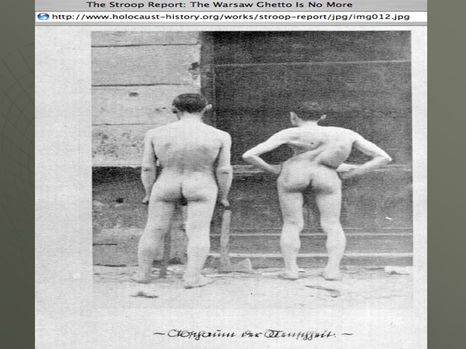
Do we have more bones when we are a baby or when we are all grown up? Baby has 305 bones and an adult has 206 bones. This is because as we grown some of our bones join together to form one bone. The longest bone in our bodies is the femur (thigh bone). The smallest bone is the stirrup bone inside the ear. Each hand has 26 bones in it. our nose and ears are not made of bone; they are made of cartilage, a flexible substance that is not as hard as bone. Differences between males and females: Males and females have slightly different skeletons, including a different elbow angle. Males have slightly thicker and longer legs and arms; females have a wider pelvis and a larger space within the pelvis, through which babies travel when they are born. Introduce some skeletal facts – could be done as a quiz to look at prior knowledge.
. The smallest bone is the stirrup bone inside the ear. Each hand has 26 bones in it. our nose and ears are not made of bone; they are made of cartilage, a flexible substance that is not as hard as bone. Differences between males and females: Males and females have slightly different skeletons, including a different elbow angle. Males have slightly thicker and longer legs and arms; females have a wider pelvis and a larger space within the pelvis, through which babies travel when they are born. Introduce some skeletal facts – could be done as a quiz to look at prior knowledge.")
5
Lets look at the skeleton
The Skeleton is the name given to the collection of bones that holds our body up. Without the trunk and branches of a tree, were would the leaves be? Without your skeleton, where would you be? Use a skeleton model, pictures and posters to introduce the skeleton to the class. Students can start thinking about why we have a skeleton.

6
Types of Skeletons Exo-skeleton: animals that have shells
Endo-skeleton: hard structure inside the animal. Hydrostatic skeleton: Fluid held inside the body No skeleton Introduce the different types of skeletons to the students. Which type do humans have? Can the students come up with other examples of these types of animals.

7
Understanding bone strength
Bone Strength Activity In your groups…….. Take a sheet of paper and curl it up. Put a piece of sticking tape on to hold it. Place as many weights on this hollow structure as possible How much weight do you think it will hold? Activity to have the students explore bone strength. The hollow paper cylinders will hold quite a lot of weight before they collapse.

8
Appendicular Skeleton
Skeletal System Function Protection Framework Movement Mineral Storage Blood Cell Formation Bone Classification Long Bones Short Bones Flat Bones Irregular Bones Composition Hard Bone Cartilage Part Appendicular Skeleton Axial Skeleton

9
Function of the Skeletal System
Support- framework that supports body and cradles its soft organs Protection- for delicate organs, heart, lungs, brain Movement- bones act as levers for muscles Mineral storage- calcium & phosphate Blood cell formation- hematopoiesis

10
The Skeletal System Parts of the skeletal system
Bones (skeleton) Joints Cartilages Ligaments (bone to bone)(tendon=bone to muscle) Divided into two divisions Axial skeleton Appendicular skeleton – limbs and girdle
 Joints. Cartilages. Ligaments (bone to bone)(tendon=bone to muscle) Divided into two divisions. Axial skeleton. Appendicular skeleton – limbs and girdle.")
11
Short Bones- carpals, tarsals Flat Bones- rib, scapula, skull, sternum
Types of Bones Long Bones- metacarples, metatarsals, phelangies, humerus, ulna, radius, tibia, fibula Short Bones- carpals, tarsals Flat Bones- rib, scapula, skull, sternum Irregular Bones- vertebrae, some facial bones Sesamoid- patella

12
Bone Classification

13
Classification of Bones
Long bones Typically longer than wide Have a shaft with heads at both ends Contain mostly compact bone Examples: Femur, humerus Copyright © 2003 Pearson Education, Inc. publishing as Benjamin Cummings

14
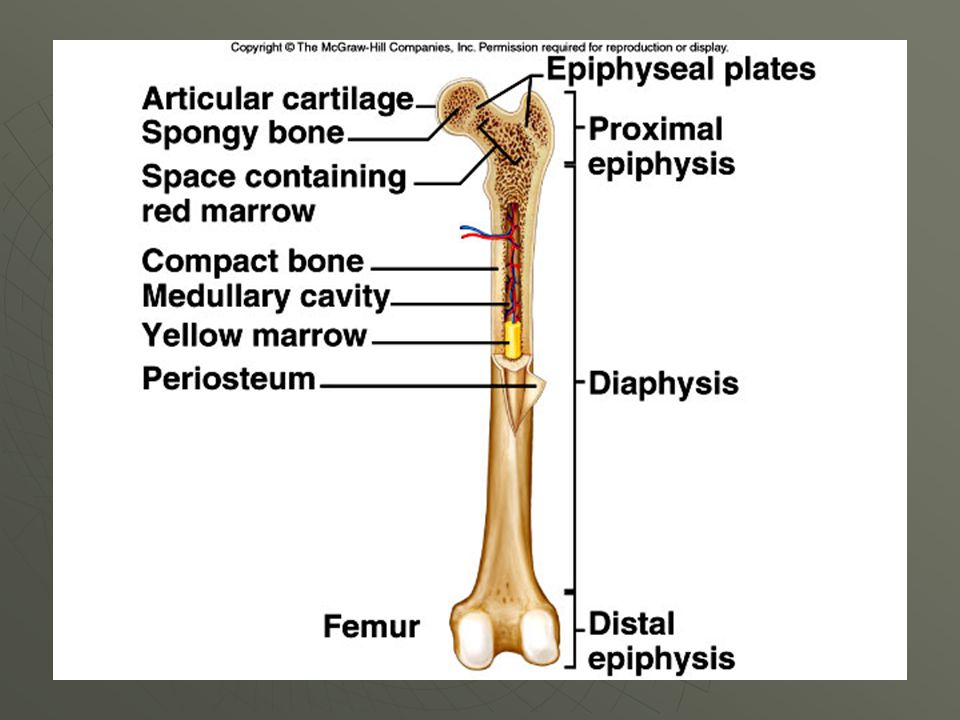
Gross Anatomy of a Long Bone
Diaphysis Shaft Composed of compact bone Epiphysis Ends of the bone Composed mostly of spongy bone Figure 5.2a Copyright © 2003 Pearson Education, Inc. publishing as Benjamin Cummings

15
275 bones 12 weeks (6-9 inches long)
Fetal Skeleton 275 bones 12 weeks (6-9 inches long)
")
16
bone cartilage calcified cartilage epiphyseal line epiphyseal plate
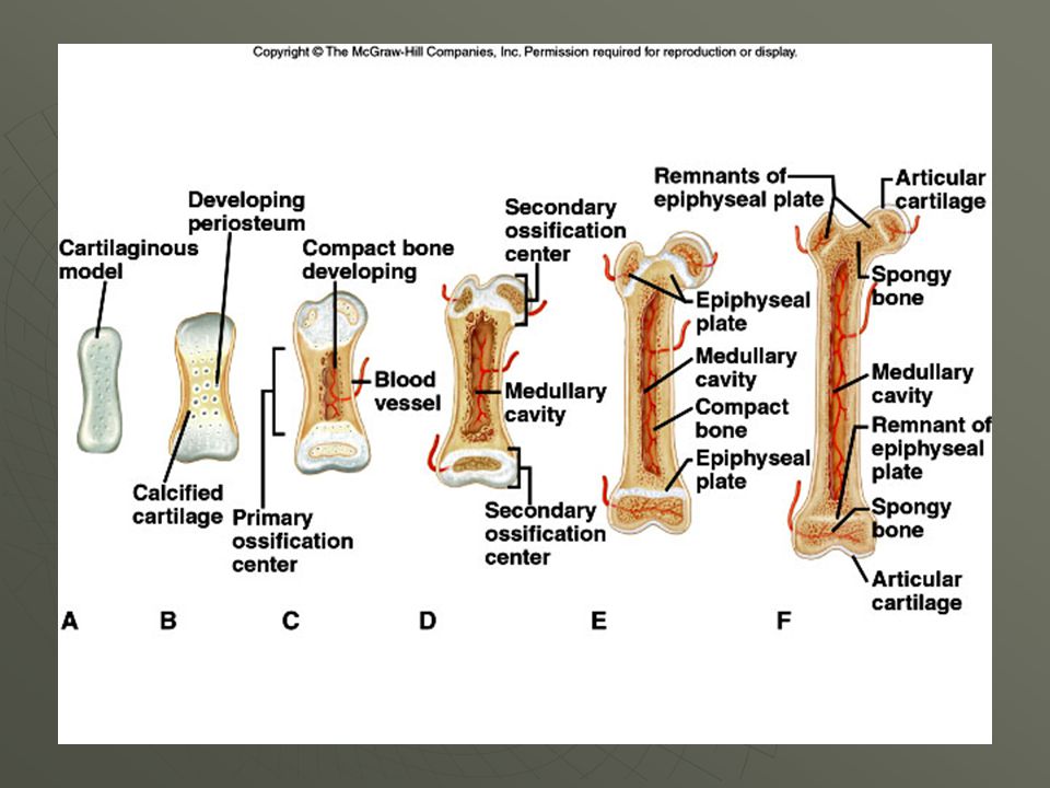
Fetus: 1st 2 months Endochondral Ossification 2o ossification center bone cartilage calcified cartilage Just before birth epiphyseal line epiphyseal plate Childhood Adult

17
Bone cells that aid in remodeling
Osteoblast Builds new bone Mature bone cell Osteocyte OsteoblastsOsteoblasts are responsible for building new bone and lie at the centre of bone physiology. Their functions include the synthesis of collagen and the control of mineralisation. OsteoclastsOsteoclasts are specialised cells that resorb bone. They work by sealing off an area of bone surface then, when activated, they pump out hydrogen ions to produce a very acid environment, which dissolves the hydroxyapatite. OsteocytesBone adapts to applied forces by growing stronger in order to withstand them; it is known that exercise can help to improve bone strength. Osteocytes are thought to be part of the cellular feed-back mechanism which directs bone to form in the places where it is most needed. They lie within mineralised bone and it is thought that they may detect mechanical deformation and mediate the response of the osteoblasts. Osteoclast Eats bone

18
Structures of a Long Bone
Periosteum Outside covering of the diaphysis Fibrous connective tissue membrane Sharpey’s fibers Secure periosteum to underlying bone Arteries Supply bone cells with nutrients Figure 5.2c Copyright © 2003 Pearson Education, Inc. publishing as Benjamin Cummings

20
Classification of Bones
Short bones Generally cube-shape Contain mostly spongy bone Examples: Carpals, tarsals Copyright © 2003 Pearson Education, Inc. publishing as Benjamin Cummings

21
Classification of Bones
Flat bones Thin and flattened Usually curved Thin layers of compact bone around a layer of spongy bone Examples: Skull, ribs, sternum Copyright © 2003 Pearson Education, Inc. publishing as Benjamin Cummings

22
Classification of Bones
Irregular bones Irregular shape Do not fit into other bone classification categories Example: Vertebrae and hip Copyright © 2003 Pearson Education, Inc. publishing as Benjamin Cummings

23
Surface features of bones
Sites of attachments for muscles, tendons, and ligaments Passages for nerves and blood vessels Categories of bone markings Projections and processes – grow out from the bone surface Depressions or cavities – indentations Copyright © 2003 Pearson Education, Inc. publishing as Benjamin Cummings

24
Sesamoid Bones Extra Bones Found in Certain Tendons i.e. Patella

25
Types of Bone Cells Osteocytes Osteoblasts Osteoclasts
Mature bone cells Osteoblasts Bone-forming cells Osteoclasts Bone-destroying cells Break down bone matrix for remodeling and release of calcium Bone remodeling is a process by both osteoblasts and osteoclasts Copyright © 2003 Pearson Education, Inc. publishing as Benjamin Cummings

26
Types of Bone Cells Osteocytes Osteoblasts Osteoclasts
Mature bone cells Osteoblasts Bone-forming cells Osteoclasts Bone-destroying cells Break down bone matrix for remodeling and release of calcium Bone remodeling is a process by both osteoblasts and osteoclasts Copyright © 2003 Pearson Education, Inc. publishing as Benjamin Cummings

27
Changes in the Human Skeleton
In embryos, the skeleton is primarily hyaline cartilage During development, much of this cartilage is replaced by bone Cartilage remains in isolated areas Bridge of the nose Parts of ribs Joints Copyright © 2003 Pearson Education, Inc. publishing as Benjamin Cummings

28
Changes in the Human Skeleton
Initially collagen fibers secreted by fibroblasts Cartilage deposited between the fibers Skeleton fully formed by 2nd month of fetal development (all cartilage) Ossification begins after 8th week of fetal development Copyright © 2003 Pearson Education, Inc. publishing as Benjamin Cummings
 Ossification begins after 8th week of fetal development. Copyright © 2003 Pearson Education, Inc. publishing as Benjamin Cummings.")
29
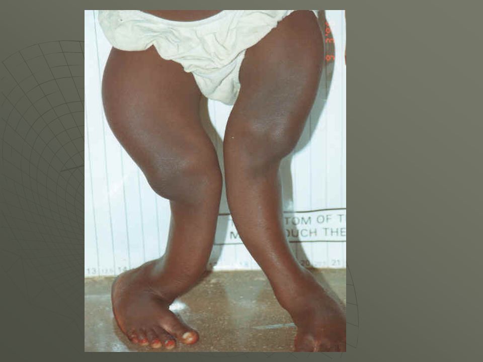
Bone Growth Epiphyseal plates allow for growth of long bone during childhood New cartilage is continuously formed Older cartilage becomes ossified Cartilage is broken down Bone replaces cartilage Copyright © 2003 Pearson Education, Inc. publishing as Benjamin Cummings

31
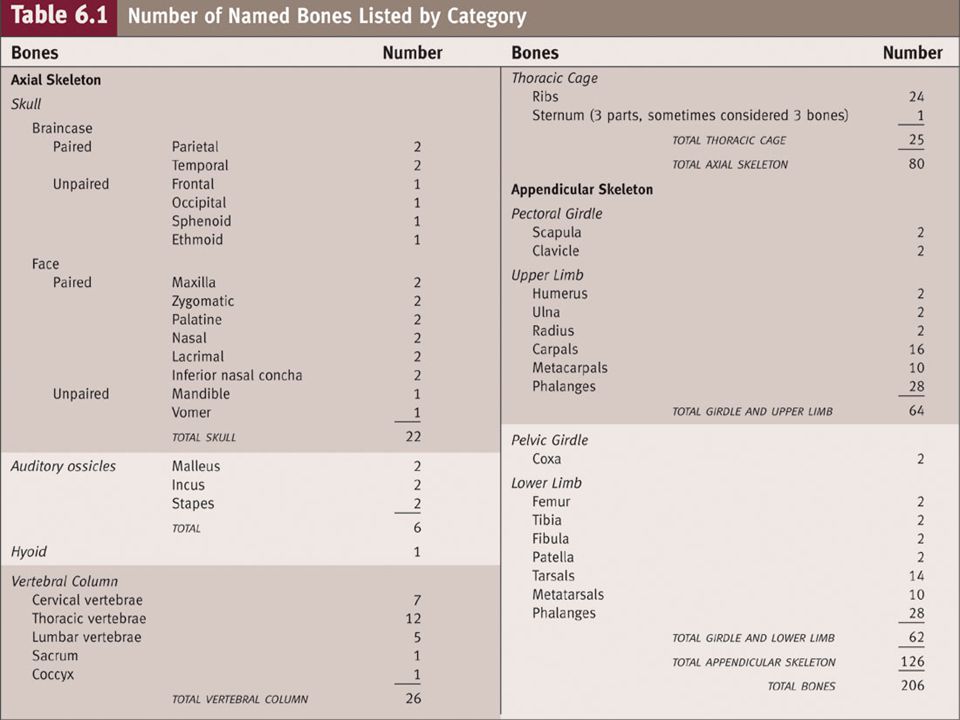
Bones of the Human Body The skeleton has 206 bones
Two basic types of bone tissue Compact bone Homogeneous Spongy bone Small needle-like pieces of bone Many open spaces Figure 5.2b Copyright © 2003 Pearson Education, Inc. publishing as Benjamin Cummings

32
Microscopic Anatomy of Bone
Figure 5.3 Copyright © 2003 Pearson Education, Inc. publishing as Benjamin Cummings

33
Based on the substance composition, the bone can be divided into hard bone and cartilage.
Based on the characteristic of its motion, articulation is difference as sinartrosis (dead joints)=sutura amfiartrosis (stiff joints)= columnar vertebrate diartrosis (joint motion)= the joint between femur and tibia
=sutura. amfiartrosis (stiff joints)= columnar vertebrate. diartrosis (joint motion)= the joint between femur and tibia.")
34
Relations between the bones with one another are called articulation or joint.
According to the direction of motion, joint is distinguished as bullets joint=shoulder with upper arm hinge joints=elbow or knees swivel joints=atlas bone with axis bone saddle joints=finger and palm sliding joints=palm and sool joints kondiloid= hand wrist

36
Based on the substance composition, the bone can be divided into hard bone and cartilage.
Based on the characteristic of its motion, articulation is difference as sinartrosis (dead joints)=sutura amfiartrosis (stiff joints)= columnar vertebrate diartrosis (joint motion)= the joint between femur and tibia
=sutura. amfiartrosis (stiff joints)= columnar vertebrate. diartrosis (joint motion)= the joint between femur and tibia.")
37
Relations between the bones with one another are called articulation or joint.
According to the direction of motion, joint is distinguished as bullets joint=shoulder with upper arm hinge joints=elbow or knees swivel joints=atlas bone with axis bone saddle joints=finger and palm sliding joints=palm and sool joints kondiloid= hand wrist

38
MATERI STRUKTUR DAN FUNGSI TULANG STRUKTUR DAN FUNGSI OTOT

39
STRUKTUR DAN FUNGSI TULANG
Sebagai pembentuk rangka tubuh Sebagai sistem gerak pasif karena adanya sendi Sebagai penyimpan Kalsium, Fosfor, Natrium dan elemen lain Sebagai penghasil sel-sel darah Proteksi terhadap organ tubuh yang lemah Pembentuk Limfosit B sebagai sistem immunologis tubuh LANJUT

40
STRUKTUR DAN FUNGSI TULANG
Rangka manusia terdiri atas RANGKA AKSIAL dan RANGKA APENDIKULER TENGKORAK = 28 BUAH TULANG SERVIK = 7 BUAH TULANG TORAKAL = 12 BUAH TULANG LUMBAL = 5 BUAH TULANG SAKRUM = 5 BUAH TULANG KOKSIGEA = 4 BUAH TULANG RUSUK SEJATI = 7 PS TULANG RUSUK PALSU = 3 PS TULANG RUSUK MELAYANG =2 PS TULANG RUSUK MELAYANG TULANG DADA = 3 BUAH Kembali RANGKA AKSIAL LANJUT

41
STRUKTUR DAN FUNGSI TULANG
Rangka Apendikuler atau rangka tam-bahan Merupakan rangka pendukung gerak/lokomosi. Terdiri atas Tungkai Atas dan Tungkai Bawah TUNGKAI ATAS TUNGKAI BAWAH RANGKA APENDIKULER KEMBAL

42
STRUKTUR DAN FUNGSI TULANG BAGIAN-BAGIAN TUNGKAI ATAS
Klavikula = 1 ps Skapula = 1 ps 1 Humerus = 1 ps 2 Ulna = 1 ps 3 Radius = 1 ps 4 Carpal = 1 ps 5 Metacarpal = 4 ps 6 7 Phalanges = 5 ps 8 KEMBAL

43
STRUKTUR DAN FUNGSI TULANG BAGIAN-BAGIAN TUNGKAI BAWAH
KOKSEA = 1 PASANG PETALA = 1 PASANG FEMUR = 1 PASANG FIBULA = 1 PASANG 1 TIBIA = 1 PASANG 2 TARSAL = 1 PASANG 3 4 METATARSAL = 4 PASANG 5 PALANGES = 5 PASANG 6 7 8 KEMBALI

44
BAGIAN–BAGIAN PENYUSUN TULANG
Sumber : 3 Endosteum Garis Epipise 2 Tulang Kompak Epipise Proksimal 1 1 2 4 Sumsum Kuning Sumsum Merah 3 Tulang Spon Periosteum 1 Tulang Kompak 2 Diapisis 2 4 Arteri Tulang Spon 3 5 1 Medula Cavity Periosteum Garis Epipise 6 Episise Distal 3

45
STRUKTUR DAN FUNGSI TULANG
BAGIAN – BAGIAN TULANG TENGKORAK Sumber : TULANG-TULANG TENGKORAK KEMBAL

46
STRUKTUR DAN FUNGSI TULANG
Bagian Penyusun Tulang Dada Sumber : Penghubung Klaikula Jugular Jugular 1 2 3 Manubrium Sternal Angle 4 Tautan rusuk 7 5 Badan Kipoid 6 KEMBAL

47
STRUKTUR DAN FUNGSI OTOT
Sebagai alat gerak aktif karena memiliki kemampuan Kontraksi, Ektensi dan Relaksasi Sebagai Penyimpan gula dalam bentuk glikogen otot MACAM OTOT STRUKTUR OTOT KONTRAKSI OTOT ANIMASI KONTRAKSI OTOT ANIMASI AKTIVASI OTOT Kembali

48
STRUKTUR DAN FUNGSI OTOT
MACAM OTOT MACAM OTOT OTOT POLOS OTOT RANGKA OTOT JANTUNG Ciri dan fungsi a. Fusiform shape b. Involuntary One nuclei in the centre Ciri dan fungsi a. voluntary b. Has not branch c. Composed of miofibril Ciri dan fungsi a. involuntary b. Has branch c. Nuclei located in the middle Kembali

49
STRUKTUR DAN FUNGSI OTOT
STRUKTUR OTOT Tendon Selaput otot Epimisium Fasikulus Endomisium Sarkolema Sarkoplasma Mio fibril Perimisium Serabut Otot Nukleus Kembali

50
STRUKTUR DAN FUNGSI OTOT
KONTRAKSI OTOT aktin Miosin Otot Berektensi Zona H memanjang Zona I memanjang Band A melebar Lanjut Miosin aktin Otot Relaksasi Zona H normal Zona I normal Band A normal Lanjut Miosin aktin Otot Berkontraksi Zona H menyempit Zona I memendek Band A memendek Kembali

Presentasi serupa