Upload presentasi
Presentasi sedang didownload. Silahkan tunggu

1
Physiology of Muscle Humaryanto

2
TIPE OTOT Otot Skeletal (lurik/striata) Otot Jantung (lurik/striata)
Otot Polos (polos) (GI, VU, Vascular)
 (GI, VU, Vascular)")
3
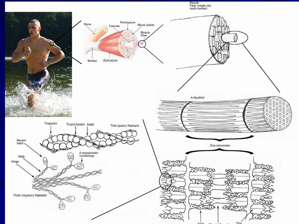
Extrafusal Muscle Fibers
Striate muscle Force for limb movements flexion - closes joint extension - opens joint Contract or relax ~

4
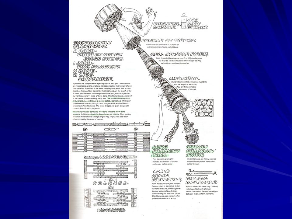
OTOT SKELETAL 40% BB tubuh Fungsi : mengatur posisi dan gerak rangka
Melekat ke tulang melalui tendo Origo : perlekatan pada bag. proksimal, bersifat stasioner Insersio : perlekatan pada bag. distal, bersifat mobil

6
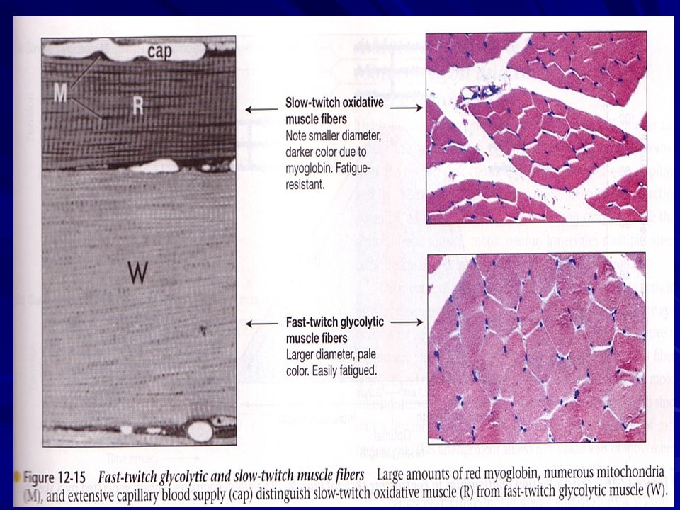
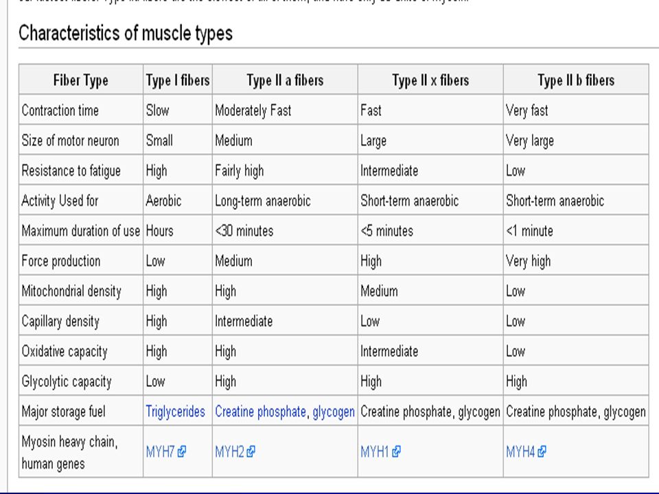
TIPE OTOT SKELETAL Berdasarkan kecepatan kontraksi dan daya tahan terhadap fatigue. Fast-twitch glycolitic fibers (putih) Fast-twitch oxidative fibers (merah) Slow twitch oxidative fibers (merah) Setiap orang punya 3 tipe otot, tapi berbeda pada komposisi dominan (Jauhari Johan vs John Murray/ Ben Johnson)
 Slow twitch oxidative fibers (merah) Setiap orang punya 3 tipe otot, tapi berbeda pada komposisi dominan. (Jauhari Johan vs John Murray/ Ben Johnson)")
7
Type 1 Fibers Slow fibers Aerobic metabolism
dark red slow, sustained contraction slow to fatigue Aerobic metabolism many capillaries & mitochondria oxygen required for ATP synthesis myoglobin gives dark red appearance ~

8
Type 2b Fibers Fast fatigable fibers Anaerobic metabolism
white fibers rapid, brief contraction fast to fatigue produce about 10x force of Type 1 Anaerobic metabolism fewer capillaries & mitochondria ATP generated by glycolysis lactic acid buildup ~

9
Type 2a Fibers Fast fatigue-resistant fibers
pale red properties intermediate to types 1 & 2b rapid, brief contraction slow to fatigue produce least force Aerobic & Anaerobic metabolism many capillaries & mitochondria ~

12
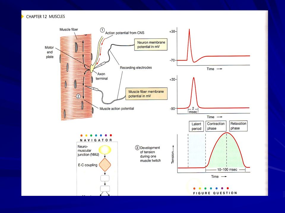
Neuromuscular Junction
Synapse between neuron & effector Cholinergic (ACh) nicotinic receptors Motor end-plate postsynaptic membrane folds packed with receptors increased surface area ~
 nicotinic receptors. Motor end-plate. postsynaptic membrane. folds packed with receptors. increased surface area ~")
13
Global view of a neuromuscular junction: 1. Axon 2. Motor end-plate 3
Global view of a neuromuscular junction: 1. Axon 2. Motor end-plate 3. Muscle fiber 4. Myofibril

14
Detailed view of a neuromuscular junction: 1. Presynaptic terminal 2
Detailed view of a neuromuscular junction: 1. Presynaptic terminal 2. Sarcolemma 3. Synaptic vesicle 4. Nicotinic acetylcholine receptor 5. Mitochondrion

15
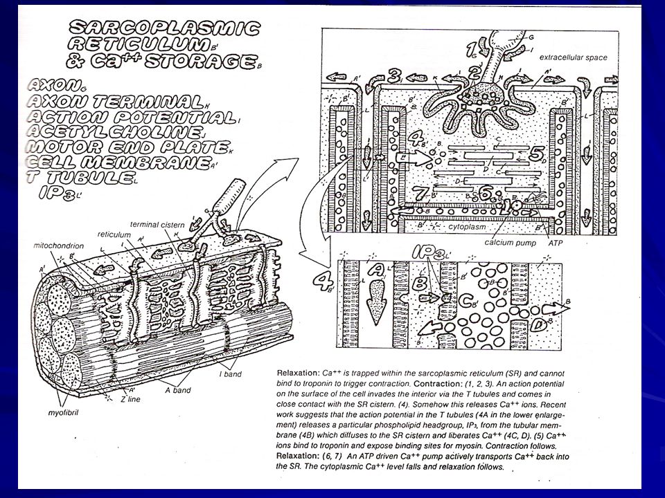
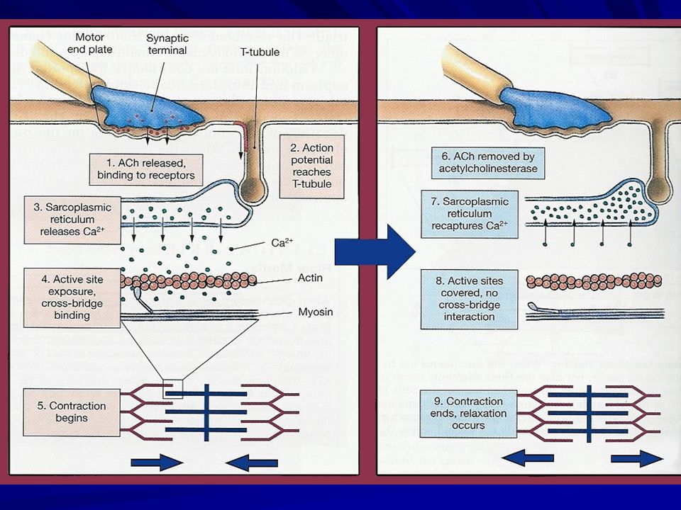
Mechanism of action Upon the arrival of an action potential at the axon terminal, voltage-dependent calcium channels open and Ca2+ ions flow from the extracellular fluid into the motor neuron's cytosol. This influx of Ca2+ triggers excitation-contraction coupling, a biochemical cascade that causes neurotransmitter-containing vesicles to fuse to the motor neuron's cell membrane and release acetylcholine into the synaptic cleft. Acetylcholine diffuses across the synaptic cleft and binds to the nicotinic acetylcholine receptors that dot the motor end plate. The receptors are ligand-gated ion channels, and when bound by acetylcholine, they open, allowing sodium and potassium ions to flow in and out of the muscle's cytosol, respectively.

16
Mechanism of action Because of the differences in electrochemical gradients across the plasma membrane, more sodium moves in than potassium out, producing a local depolarization of the motor end plate known as an end-plate potential (EPP). This depolarization spreads across the surface of the muscle fiber into transverse tubules, eliciting the release of calcium from the sarcoplasmic reticulum, thus initiating muscle contraction. The action of acetylcholine is terminated when the enzyme acetylcholinesterase degrades the neurotransmitter and the unhydrolysed neurotransmitter diffuses away
. This depolarization spreads across the surface of the muscle fiber into transverse tubules, eliciting the release of calcium from the sarcoplasmic reticulum, thus initiating muscle contraction. The action of acetylcholine is terminated when the enzyme acetylcholinesterase degrades the neurotransmitter and the unhydrolysed neurotransmitter diffuses away.")
17
Neurotransmitters and Neuromodulators
Neuromodulators modify the postsynaptic cell's response to neurotransmitters or change the presynaptic cell's synthesis, release or metabolism of the neurotransmitter. Acetylcholine (Ach) Major neurotransmitter. Fibers that release ACh are called cholinergic fibers. Acetylcholine is degraded by the enzyme, acetylcholinesterase. Biogenic Amines Biogenic amines are neurotransmitters containing an amino group. Catecholamines such as dopamine, norepinephrine and epinephrine, serotonin. Nerve fibers that release epinephrine and norepinephrine are called adrenergic and noradrenergic fibers respectively.
 Major neurotransmitter. Fibers that release ACh are called cholinergic fibers. Acetylcholine is degraded by the enzyme, acetylcholinesterase. Biogenic Amines. Biogenic amines are neurotransmitters containing an amino group. Catecholamines such as dopamine, norepinephrine and epinephrine, serotonin. Nerve fibers that release epinephrine and norepinephrine are called adrenergic and noradrenergic fibers respectively.")
18
Neurotransmitters and Neuromodulators
Amino Acid Neurotransmitters Amino acid neurotransmitters are the most prevalent neurotransmitters in CNS. Glutamate, aspartate GABA (gamma aminobutyric acid), glycine, Neuropeptides Neuropeptides are composed of two or more amino acids. Neurons releasing neuropeptides are called peptidergic. Beta-endorphin, dynorphin, enkephalins. Nitric oxide, ATP, adenine also act as neurotransmitters. Neuroeffector Communication Many neurons of peripheral nervous system end at neuroeffector junctions on muscle and gland cells. Neurotransmitters released by these efferent neurons then activate the target cell.
, glycine, Neuropeptides. Neuropeptides are composed of two or more amino acids. Neurons releasing neuropeptides are called peptidergic. Beta-endorphin, dynorphin, enkephalins. Nitric oxide, ATP, adenine also act as neurotransmitters. Neuroeffector Communication. Many neurons of peripheral nervous system end at neuroeffector junctions on muscle and gland cells. Neurotransmitters released by these efferent neurons then activate the target cell.")
19
Muscle Contraction AP generated in muscle fiber (cell)
Ca++ released from internal stores Muscle fiber contracts continues while Ca++ & ATP available Relaxation Ca++ sequestered by active transport ~

20
Muscle Fiber Structure
Multinucleated fusion of multiple precursor cells Sarcolemma Excitable membrane Myofibrils: contractile units Sarcopasmic reticulum (SR) sequesters Ca++ T tubules AP from sarcolemma to SR like inside-out axons ~
 sequesters Ca++ T tubules. AP from sarcolemma to SR. like inside-out axons ~")
21
Miofibril struktur kontraksi otot
1 Serat otot, tdd: ribuan miofibril 1 miofibril tdd: Aktin & miosin (protein kontraksi) Troponin & tropomiosin (protein pengatur) Titin & nebulin (protein asessoris besar) Miosin thick filament, punya kepala Motor protein, E kimia E mekanik, mgd ATP-ase (hidrolisis) Aktin thin filament, melekat troponin & tropomiosin Titin molekul elastis (protein terbesar) stabilitas & elastisistas otot Nebulin penyanggah aktin
 Troponin & tropomiosin (protein pengatur) Titin & nebulin (protein asessoris besar) Miosin thick filament, punya kepala. Motor protein, E kimia E mekanik, mgd ATP-ase (hidrolisis) Aktin thin filament, melekat troponin & tropomiosin. Titin molekul elastis (protein terbesar) stabilitas & elastisistas otot. Nebulin penyanggah aktin.")
22
Sarcoplasmic Reticulum T tubules Myofibrils Sarcolemma

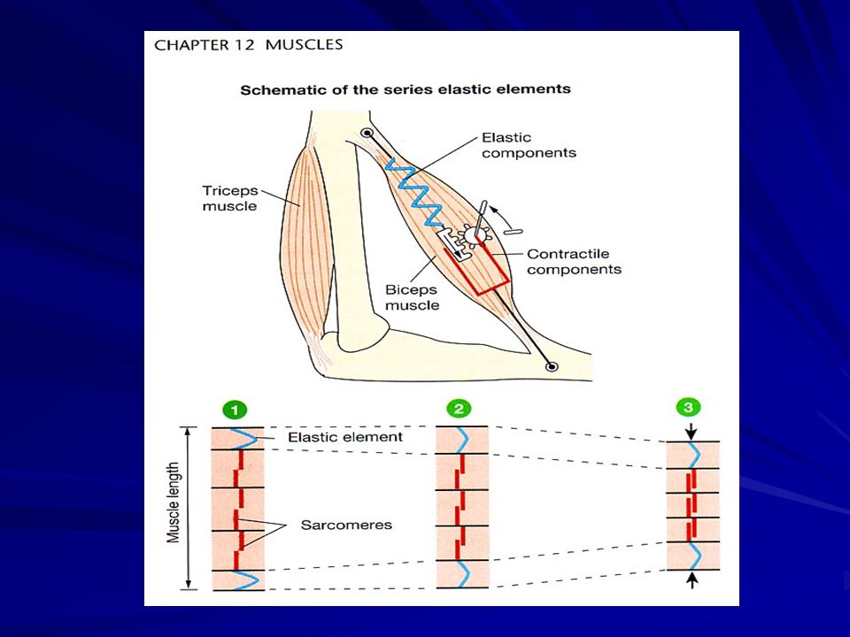
24
Myofibril: structure & function
Sarcomeres repeating sections Z lines dividers between sarcomeres thin filaments anchored to Z lines actin & troponin Thick filaments between thin filaments myosin Contraction:filaments slide by each other ~

25
Z line Thin filaments Thick Filaments Sarcomere

27
KONTRAKSI OTOT Menghasilkan force / gaya muscle tension
Melawan beban/ load Memerlukan energi (dari ATP) Pencetus kontraksi otot 1. Neuromuscular junction : Rangsang somatik rangsang listrik 2. Excitation-contraction coupling Potensial aksi signal Ca++ siklus kontr-relaks SIKLUS KONTRAKSI DAN RELAKSASI Sliding filaments theory
 Pencetus kontraksi otot. 1. Neuromuscular junction : Rangsang somatik rangsang listrik. 2. Excitation-contraction coupling. Potensial aksi signal Ca++ siklus kontr-relaks. SIKLUS KONTRAKSI DAN RELAKSASI. Sliding filaments theory.")
28
Contraction Excitation-contraction coupling
Myosin “heads” crossbridges w/ actin Ca++ dependent binds to troponin, reveals binding site Myosin head rotates “ratchets” actin inward ~

29
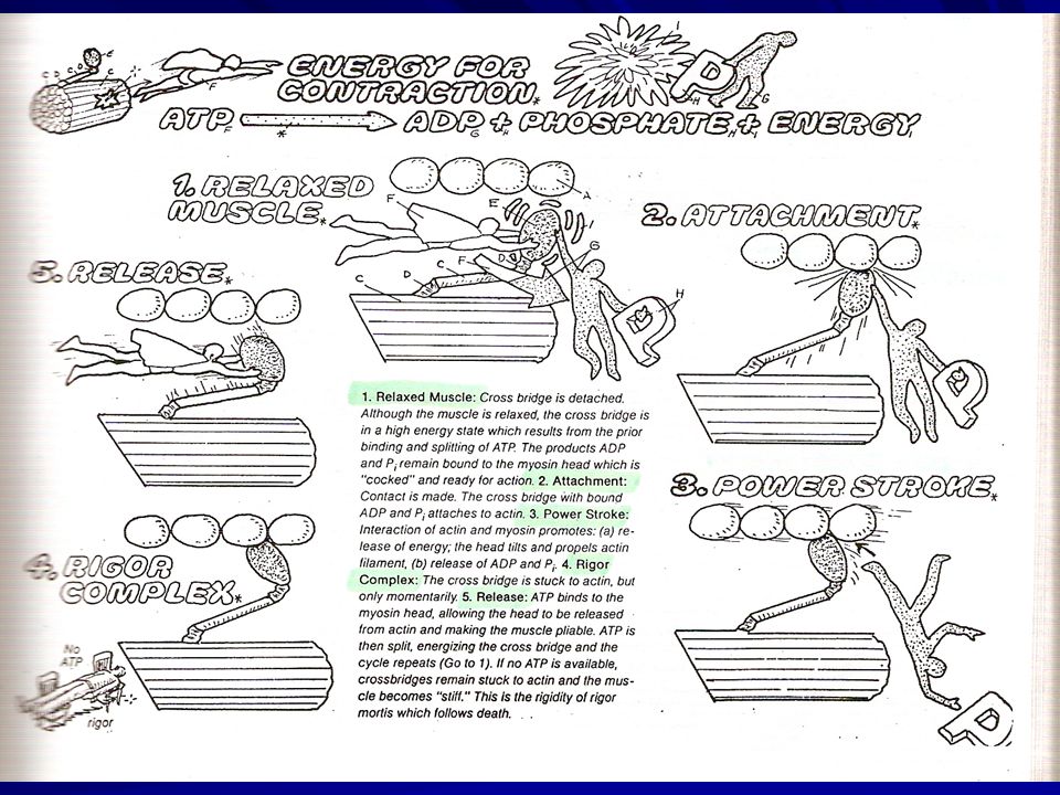
Contraction ATP binds to myosin ---> detachment
cocks myosin ---> binds again rigor mortis: no ATP fibers remain crosslinked Repeats as long as Ca++ present sequestered via active transport ~

30
SLIDING FILAMENT THEORY
Serat otot memendek (overlapping thick & thin filament) Sliding aktin terhadap miosin Gaya dari crossbridge miosin mendorong aktin (power stroke) Crossbridge miosin mendorong aktin menuju pusat sarkomer Setelah power stroke kepala miosin melepas aktin untuk mengikat bagian aktin yang lain, demikian seterusnya jadi siklus. Analogi : menarik tambang.
 Sliding aktin terhadap miosin. Gaya dari crossbridge miosin mendorong aktin. (power stroke) Crossbridge miosin mendorong aktin menuju pusat sarkomer. Setelah power stroke kepala miosin melepas aktin untuk mengikat bagian aktin yang lain, demikian seterusnya jadi siklus. Analogi : menarik tambang.")
32
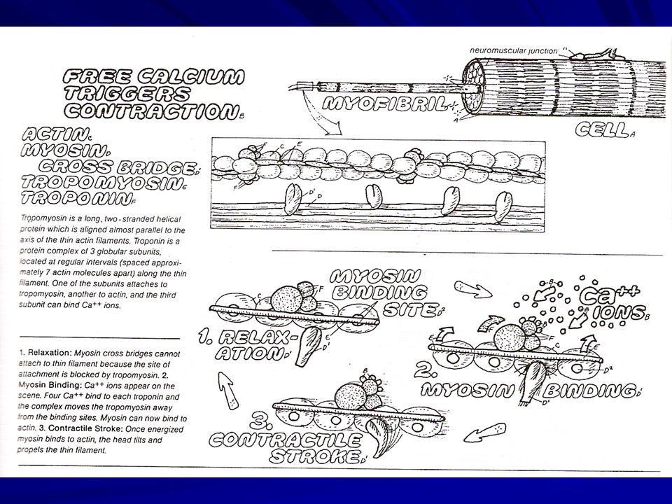
In the absence of calcium ions, tropomyosin blocks access to the mysosin binding site of actin.
When calcium binds to troponin, the positions of troponin and tropomyosin are altered on the the thin flament and myosin then has access to its binding site on actin. Myosin hydolyzes ATP and undergoes a conformational change into a high-energy state. The head group of myosin binds to actin forming a cross-bridge between the thick and thin filaments.

33
Role of Ca+2 in Muscle Contraction
* Actin-binding sites are exposed as a result of Ca+2 binding to troponin complex that causes a conformational shift of tropomyosin

35
The energy stored by myosin is released, and ADP and inorganic phosphate dissociate from myosin.
The resulting relaxation of the myosin molecule entails rotation of the globular head, which induces longitudinal sliding of the filaments. When the calcium level decreases, troponin locks tropomyosin in the blocking position and the thin filament slides back to the resting state.

37
Sliding-Filament Mechanism
Muscle contraction is produced by cross bridge cycles. A cycle has 4 steps: (1) Energizing of myosin cross bridge A + M•ATP —> A + M*•ADP•Pi (ATP is energizer here) (2) Attachment of cross bridge to a thin filament A + M*•ADP•Pi —> A•M*•ADP•Pi (3) Movement of cross bridge, producing tension A•M*•ADP•Pi —> A•M + ADP + Pi (4) Detachment of cross bridge from thin filament A•M + ATP —> A + M•ATP (ATP is modulator here) Movement of the cross bridges make the overlapping thick and thin filaments slide past each other (they do not change in length) to produce a contraction.
 Energizing of myosin cross bridge. A + M•ATP —> A + M*•ADP•Pi (ATP is energizer here) (2) Attachment of cross bridge to a thin filament. A + M*•ADP•Pi —> A•M*•ADP•Pi. (3) Movement of cross bridge, producing tension. A•M*•ADP•Pi —> A•M + ADP + Pi. (4) Detachment of cross bridge from thin filament. A•M + ATP —> A + M•ATP (ATP is modulator here) Movement of the cross bridges make the overlapping thick and thin filaments slide past each other (they do not change in length) to produce a contraction.")
38
Actin Myofilament During contraction, calcium binds to troponin
Covers actin-binding sites at rest

39
Cross-Bridge Formation

40
Cross-Bridge Cycle

41
Cross-bridge Cycle This animation by Mike Geeves, Laboratory of Molecular Biology in the UK and the Cambridge Institute for Medical Research

42
SIKLUS KONTRAKSI Rigor state: Kepala miosin terikat dg molekul G-aktin. ATP menempel ke miosin, kepala miosin lepas dari aktin. Hidrolisis ATP: jadi ADP + Pi (masih menempel) Miosin melekat ke G-aktin yang baru, energi dari pecahnya ATP, saat ada potensial energi di kepala miosin untuk power stroke. Pi lepas & power stroke: Kepala miosin berotasi mendorong aktin mendekati pusat sarkomer (crossbridge tilting) ADP lepas: kepala miosin tetap melekat ke aktin, siap untuk siklus berikut bila ada ATP yang baru
 Miosin melekat ke G-aktin yang baru, energi dari pecahnya ATP, saat ada potensial energi di kepala miosin untuk power stroke. Pi lepas & power stroke: Kepala miosin berotasi mendorong aktin mendekati pusat sarkomer (crossbridge tilting) ADP lepas: kepala miosin tetap melekat ke aktin, siap untuk siklus berikut bila ada ATP yang baru.")
43
Excitation-Contraction Coupling

44
Excitation-Contraction Coupling
Excitation-Contraction (EC) Coupling: 1. An AP travels down a motor (somatic neuron). 2. The AP causes the release of the neurotransmitter acetylcholine into the synapse at the neuromuscular junction. 3. The acetylcholine binds to the acetylcholine receptors on the muscle fiber and cause an EPSP. 4. If the EPSP reaches threshold, an AP is produced on the sarcolemma of the muscle fiber. Meanwhile, the acetylcholine attached to the receptor is destroyed. 5. The AP travels rapidly along the sarcolemma and enters the fiber at every t-tubule.
 Coupling: 1. An AP travels down a motor (somatic neuron). 2. The AP causes the release of the neurotransmitter acetylcholine into the synapse at the neuromuscular junction. 3. The acetylcholine binds to the acetylcholine receptors on the muscle fiber and cause an EPSP. 4. If the EPSP reaches threshold, an AP is produced on the sarcolemma of the muscle fiber. Meanwhile, the acetylcholine attached to the receptor is destroyed. 5. The AP travels rapidly along the sarcolemma and enters the fiber at every t-tubule.")
45
Excitation-Contraction Coupling
6. As the AP travels through the t-tubule, it causes the Ca++ gates to open and Ca++ flows from the SR into the sarcoplasm. The Ca++ gates close when the AP ends. 7. The increased [Ca++] in the sarcoplasm results in Ca++ binding to troponin. This induces an allosteric change, the tropomyosin is pulled out of the way and steric inhibition is removed. The result is crossbridges begin to form, rotate and break (provided there is plenty of ATP). 8. Cross-bridge cycling continues as long as sarcoplasmic [Ca++] remains high. 9. However, if the Ca++ gates close, the action of the Ca++ ATPase (pump) begins to predominate and sarcoplasmic [Ca++]] drops. When it drops low enough, the troponin loses its Ca++ and changes shape the next time a crossbridge is not in the way. Steric inhibition is quickly re-established and the muscle contraction is over.
. 8. Cross-bridge cycling continues as long as sarcoplasmic [Ca++] remains high. 9. However, if the Ca++ gates close, the action of the Ca++ ATPase (pump) begins to predominate and sarcoplasmic [Ca++]] drops. When it drops low enough, the troponin loses its Ca++ and changes shape the next time a crossbridge is not in the way. Steric inhibition is quickly re-established and the muscle contraction is over.")
47
Exitation-Contraction Coupling
Dirangsang oleh asetilkolin/achetylcholine Tahap: Asetilkolin (Ach) lepas dari motor neuron somatik Ach merangsang potensial aksi serat otot PA, m’rsg Ca++ lepas dr Ret.Sarkoplasma Ca++ me’ikat troponin dan m’rsg kontraksi
 lepas dari motor neuron somatik. Ach merangsang potensial aksi serat otot. PA, m’rsg Ca++ lepas dr Ret.Sarkoplasma. Ca++ me’ikat troponin dan m’rsg kontraksi.")
48
DHP: Dihydropiridine Saat PA: Ca 100x Relaksasi: Ca masuk RS krn enzim Ca-ATP-ase

49
PERIODE KONTRAKSI/ TWITCH
Periode Laten (Antara potensial aksi-kontraksi) 2. Periode kontraksi 3. Periode relaksasi Lama periode kontraksi tergantung tipe otot
 2. Periode kontraksi. 3. Periode relaksasi. Lama periode kontraksi tergantung tipe otot.")
51
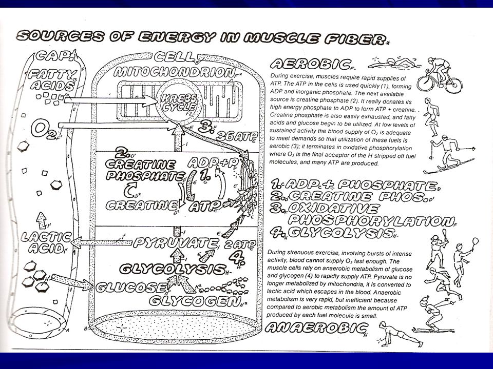
SUMBER ENERGI KONTRAKSI
ATP (Adenosine Tri Phosphate) Kontraksi: gerakan crossbridge Relaksasi: Ca++ masuk lagi ke RS Relaksasi: melepas ikatan aktin dan miosin Diluar periode kontraksi : restore Na-K SUMBER ATP Konversi posfo-kreatin (8 twitch) An-aerobik glikolisis Posforilasi-oksidatif
 Kontraksi: gerakan crossbridge. Relaksasi: Ca++ masuk lagi ke RS. Relaksasi: melepas ikatan aktin dan miosin. Diluar periode kontraksi : restore Na-K. SUMBER ATP. Konversi posfo-kreatin (8 twitch) An-aerobik glikolisis. Posforilasi-oksidatif.")
53
KELELAHAN OTOT Fatigue:
Kondisi dimana otot tidak mampu lagi melakukan / mempertahankan kontraksi Jenis Fatigue: Sentral: SSP Perifer: NM-Junction – elemen kontraksi E/ >> elemen kontraksi

54
Lelah Sentral SSP Psikologis Refleks Proteksi Asidosis (as. Laktat) NM-Junction Pelepasan Neurotransmitter dan sensitivitas reseptor Ggn. Neuromuskuler (Ach ) Kelelahan Perifer Excitation-contraction coupling Perubahan membran potensial Gangguan elektrolit (Hipokalemia) Ca++ signal Pelepasan Ca & interaksi dg troponin Pi Kontraksi-relaksasi PCr, ATP, glikogen H+, Pi, laktat
 Kelelahan. Perifer. Excitation-contraction coupling. Perubahan membran potensial. Gangguan elektrolit (Hipokalemia) Ca++ signal. Pelepasan Ca & interaksi dg troponin. Pi. Kontraksi-relaksasi. PCr, ATP, glikogen. H+, Pi, laktat.")
55
Daya kontraksi (tension) maksimal tjd pada panjang sarokomere yang optimal
 maksimal tjd pada panjang sarokomere yang optimal")
56
Tension juga meningkat bila stimulus dilakukan berulang kali sebelum mencapai relaksasi maksimum (Stimulus Summation) Akan tetapi, bila stimulus (potensial aksi) berlangsung terus menerus dg cepat (frekuensi tinggi), tanpa fase relaksasi terjadi Tetanus Tetanus Komplet/ fused Inkomplet / unfused
 berlangsung terus menerus dg cepat (frekuensi tinggi), tanpa fase relaksasi terjadi Tetanus. Tetanus. Komplet/ fused. Inkomplet / unfused.")
57
Muscle Adaptation to Exercise
Increased amount of contractile activity (exercise) increases size (hypertrophy) of muscle fibers and capacity for ATP production. Low intensity exercise affects oxidative fibers, increasing the number of mitochondria and capillaries. High intensity exercise affects glycolytic fibers, increasing their diameter by an increased synthesis of actin and myosin filaments, and an increased synthesis of glycolytic enzymes.
 increases size (hypertrophy) of muscle fibers and capacity for ATP production. Low intensity exercise affects oxidative fibers, increasing the number of mitochondria and capillaries. High intensity exercise affects glycolytic fibers, increasing their diameter by an increased synthesis of actin and myosin filaments, and an increased synthesis of glycolytic enzymes.")
58
Speed of Muscle Contraction Varies by Fiber Type

59
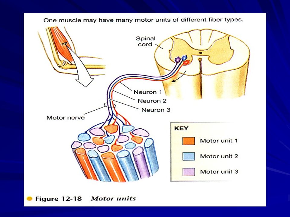
MOTOR UNIT Unit dasar kontraksi, tdd: bbrp serat otot + motor neuron somatik Motor neuron mencetuskan potensial aksi kontraksi 1 motor unit. 1 motor neuron bbrp otot; 1 serat otot dipersyarafi 1 neuron Otot kecil (gerak halus; tangan, wajah) 1 motor unit = 3-5 serat otot Otot besar (gerak kasar; tungkai, trunkus) 1 motor unit = 100an- 1000an serat otot
 1 motor unit = 3-5 serat otot. Otot besar (gerak kasar; tungkai, trunkus) 1 motor unit = 100an- 1000an serat otot.")
60
Motor Pools & Motor Units
all a motor neurons that innervate a single muscle An a motor neuron and all the muscle fibers that it innervates 1:3 to 1:100 fewer muscle fibers ---> finer control 3 types based on speed of contraction & fatigue ~

61
Types of Motor Units Most muscle contain both slow- & fast-twitch fibers ratio depends on function e.g. ankle extensors Soleus active during standing hi ratio of slow fibers Medial Gastrocnemius: active during running & jumping hi ratio of fast fibers ~

63
Variasi gradasi, gaya & durasi kontrksi ditentukan oleh :
Jumlah & jenis motor unit yang aktif (‘Recruitment’ dikontrol oleh SSP) Kontraksi lemah SSP m’rsg sedikit motor unit Motor unit yang t’rsg Ix nilai ambang rendah slow-twitch Stimulus m’rsg motor neuron dg nilai ambang tinggi fast-twitch Jumlah motor unit daya kontraksi Asynchronous Recruitment Pada kontraksi lama, SSP m’rsg bbrp motor unit scr bergantian 1 serat kontraksi, serat lain istirahat Hanya terjadi pada kontraksi sub-maksimal, why ?
 Kontraksi lemah SSP m’rsg sedikit motor unit. Motor unit yang t’rsg Ix nilai ambang rendah slow-twitch. Stimulus m’rsg motor neuron dg nilai ambang tinggi fast-twitch. Jumlah motor unit daya kontraksi Asynchronous Recruitment. Pada kontraksi lama, SSP m’rsg bbrp motor unit scr bergantian 1 serat kontraksi, serat lain istirahat. Hanya terjadi pada kontraksi sub-maksimal, why")
64
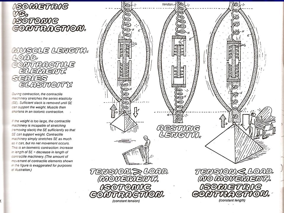
BIOMEKANIKA GERAK TUBUH
Fs otot: menggerakkan rangka KONTRAKSI ISOTONIK (Iso;=, teinein; stretch) Kontraksi gaya + menggerakkan beban Sarkomer menarik beban dan serat elastis Konsentrik arah gerak = pemendekan otot Eksentrik arah gerak = pemanjangan otot >> m’rusak otot (DOMS) KONTRAKSI ISOMETRIK (Iso;=, metric;ukuran) Kontraksi gaya + tanpa menggerakkan beban Sarkomer otot hanya menarik serat elastis
 Kontraksi gaya + menggerakkan beban. Sarkomer menarik beban dan serat elastis. Konsentrik arah gerak = pemendekan otot. Eksentrik arah gerak = pemanjangan otot. >> m’rusak otot (DOMS) KONTRAKSI ISOMETRIK (Iso;=, metric;ukuran) Kontraksi gaya + tanpa menggerakkan beban. Sarkomer otot hanya menarik serat elastis.")
68
USAHA OTOT Lever/ lengan dibentuk oleh rangka
Fulcrum/ sumbu dibentuk sendi W = F x d W (otot) = W (beban) Insersi bisep 5 cm dari siku Panjang lengan 20 cm Berat beban 5 kg Berapa usaha otot bisep mengangkat beban ? Semakin dekat insersi ke fulcrum Gerak semakin luas
 = W (beban) Insersi bisep 5 cm dari siku. Panjang lengan 20 cm. Berat beban 5 kg. Berapa usaha otot bisep mengangkat beban Semakin dekat insersi ke fulcrum. Gerak semakin luas.")
69
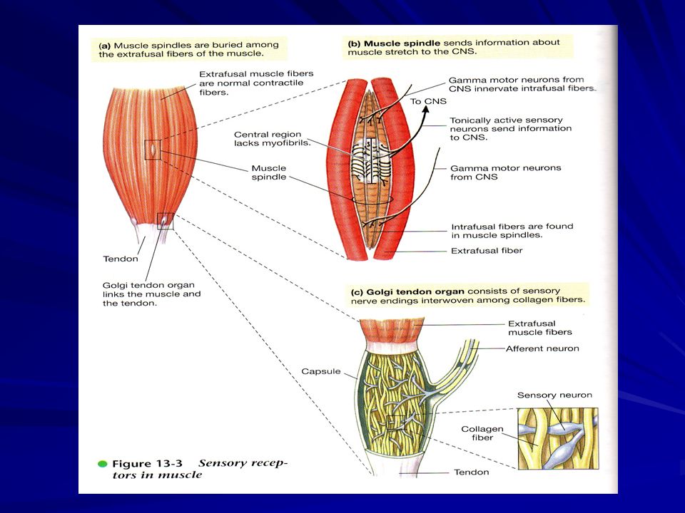
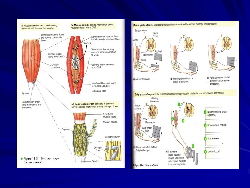
Refleks Otot Skeletal Berfungsi utk: Mengatur keseimbangan
Gerak spesifik (keselamatan) Optimalisasi gerak Komponen Refleks 1.Reseptor sensoris (proprioceptors) Spindle otot, organ tendo Golgi & reseptor sendi 2. Neuron sensoris (transfer input) 3. SSP 4. Motor neuron somatik (alfa motor neuron) 5. Serat otot (serat ekstrafusal)
 Optimalisasi gerak. Komponen Refleks. 1.Reseptor sensoris (proprioceptors) Spindle otot, organ tendo Golgi & reseptor sendi. 2. Neuron sensoris (transfer input) 3. SSP. 4. Motor neuron somatik (alfa motor neuron) 5. Serat otot (serat ekstrafusal)")
71
Reseptor Sendi Terdapat di kapsul sendi dan ligamen Distimulasi oleh distorsi mekanik krn perubahan sudut, beban dan posisi sendi & tulang Pusat pengaturan di cerebellum

72
Spindle Otot Reseptor regangan otot Mengirim impuls ke Med. Spinalis & otak Responsif thd perubahan panjang otot 1 otot memiliki bbrp spindle otot, kec, rahang Terletak di sisi dalam otot ekstrafusal, mengelilingi otot intrafusal Akibat dari rangsangan menghasilkan refleks kontraksi

73
Organ Tendo Golgi Terletak di sambungan tendo dan otot Responsif terhadap tegangan otot Menghasilkan refleks relaksasi Terdiri dari ujung syaraf bebas Refleks menghambat interneuron di MS, interneuron menghambat alfa motor neuron sehingga kontraksi berkurang.

75
Knee Jerk reflexes; merangsang spindle otot Flexion reflexes; menghindari bahaya

76
Movement Disorders of Muscle

77
Duchenne’s Muscular Dystrophy
Muscular Dystrophies wasting away of muscles metabolic / structural abnormalities Duchenne’s best understood young boys ~

78
Duchenne’s Muscular Dystrophy
Cause hereditary - maternal X chromosome single gene ---> protein dystrophin maybe involved in Ca++ regulation Treatment Inject dystrophin or mRNA Gene therapy promising for muscles ~

79
Myasthenia Gravis Severe muscle weakness
rapid fatigue following exercise Develops in people of all ages Most common: women in 30s Risk of respiratory paralysis Autoimmune disorder body develops antibodies for ACh-R reduces synaptic transmission ~

80
Myasthenia Gravis: Treatment
AChE inhibitors ¯ degradation of ACh narrow therapeutic window too much ACh ---> paralysis Reduce immune response remove thymus filtering antibodies from blood temporary ~

Presentasi serupa


>")

 REPETITIVE NERVE STIMULATION (Electromyography & Neuromuscular Disorders,>")


 Fusiform (parallel)>")


