Upload presentasi
Presentasi sedang didownload. Silahkan tunggu

1
Bases and nucleotides The nucleic acids play a central role in the storage and expression of genetic information. They are divided into two major classes: (1)deoxyribonucleic acid (DNA) functions solely in information storage, (2)ribonucleic acids (RNAs) are involved in most steps of gene expression and protein biosynthesis. All nucleic acids are made up from nucleotide components, consist of a base, a sugar, and a phosphate residue
deoxyribonucleic acid (DNA) functions solely in information storage, (2)ribonucleic acids (RNAs) are involved in most steps of gene expression and protein biosynthesis. All nucleic acids are made up from nucleotide components, consist of a base, a sugar, and a phosphate residue.")
2
GULA RIBOSA Gula pada asam nukleat adalah ribosa.
Ribosa (b-D-furanosa) adalah gula pentosa (jumlah karbon 5). Perhatikan penomoran. Dalam penulisan diberi tanda prime(') untuk membedakan penomoran pada basa nitrogen 5 1 2 3 4 KRT-2011
 adalah gula pentosa (jumlah karbon 5). Perhatikan penomoran. Dalam penulisan diberi tanda prime( ) untuk membedakan penomoran pada basa nitrogen KRT")
3
PERHATIKAN Ikatan gula ribosa dengan basa nitrogen (pada atom karbon nomor 1). Ikatan gula ribosa dengan gugus fosfat (pada atom karbon nomor 5). Gugus hidroksil pada atom karbon nomor 2 KRT-2011

4
BASA NITROGEN Basa nitrogen berikatan dengan ikatan-b pada atom karbon nomor1' dari gula ribosa atau deoksiribosa. Pirimidin berikatan ke gula ribosa pada atom N-1 dari struktur cincinnya. Purin berikatan ke gula ribosa pada atom N-9 dari struktur cincinnya. KRT-2011

5
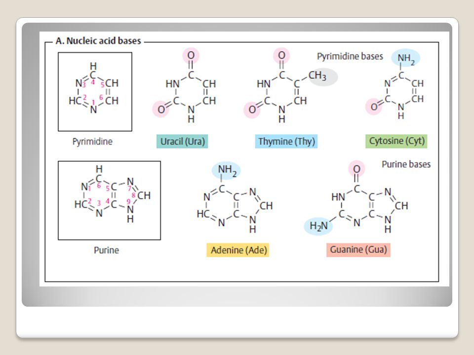
BASA PIRIMIDIN DAN PURIN
Perhatikan struktur cincinnya KRT-2011

6
GUGUS FOSFAT Nukleosida (Gula Ribosa yang berikatan dengan basa nitrogen) + satu atau lebih gugus fosforil disebut nukleotida. KRT-2011

7
Nucleic acid bases The bases that occur in nucleic acids are aromatic heterocyclic compounds derived from either pyrimidine or purine. The purine bases adenine (abbreviation Ade, not “A”!) and guanine (Gua) and the pyrimidine base cytosine (Cyt) are present in both RNA and DNA. In contrast, uracil (Ura) is only found in RNA. In DNA, uracil is replaced by thymine (Thy),
 and guanine. (Gua) and the pyrimidine base cytosine. (Cyt) are present in both RNA and DNA. In. contrast, uracil (Ura) is only found in RNA. In. DNA, uracil is replaced by thymine (Thy),")
9
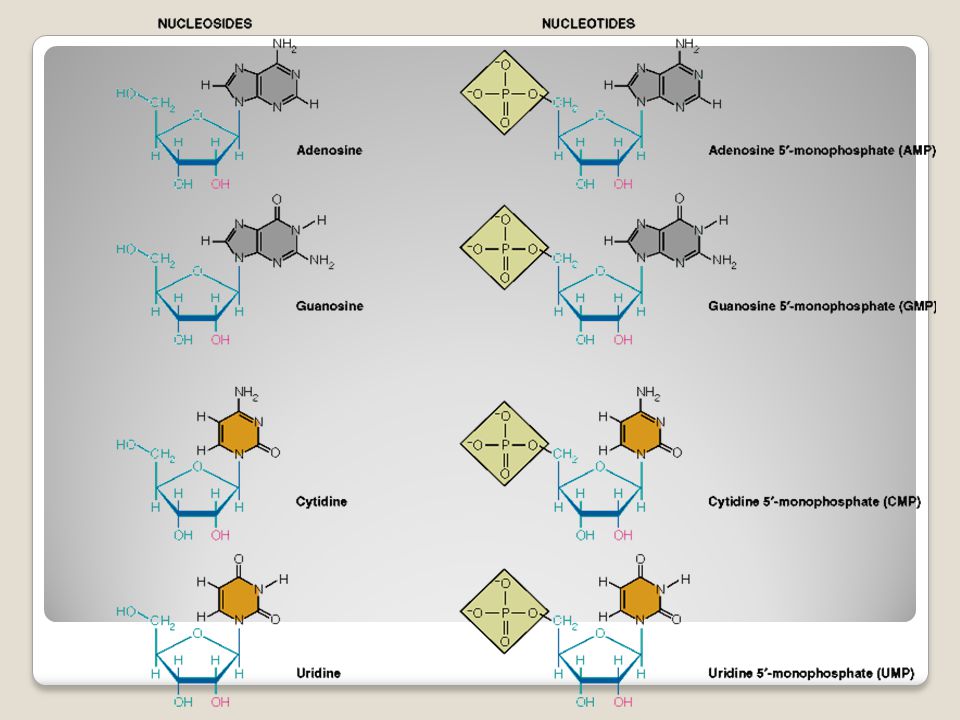
When a nucleic acid base is N-glycosidically linked to ribose or 2- deoxyribose, it yields a nucleoside. The nucleoside adenosine (abbreviation: A) is formed in this way from adenine and ribose, for example. The corresponding derivatives of the other bases are called guanosine (G), uridine (U), thymidine (T) and cytidine (C). When the sugar component is 2-deoxyribose, the product is a deoxyribonucleoside—e. g., 2-deoxyadenosine (dA, not shown).
 is formed in this way from adenine and ribose, for example. The corresponding derivatives of the other bases are called guanosine (G), uridine (U), thymidine (T) and cytidine (C). When the sugar component is 2-deoxyribose, the product is a deoxyribonucleoside—e. g., 2-deoxyadenosine (dA, not shown).")
10
In the cell, the 5OH group of the sugar component of the nucleoside is usually esterified with phosphoric acid. If the 5phosphate residue is linked via an acid–anhydride bond to additional phosphate residues, it yields nucleoside diphosphates and triphosphates—e. g., ADP and ATP, which are important coenzymes in energy metabolism. All of these nucleoside phosphates are classified as nucleotides.

11
Polynucleotides consisting of ribonucleotide components are called ribonucleic acid (RNA), while those consisting of deoxyribo-nucleotide monomers are called deoxyribonucleicacid (DNA).
, while those consisting of deoxyribo-nucleotide monomers are called deoxyribonucleicacid (DNA).")
12
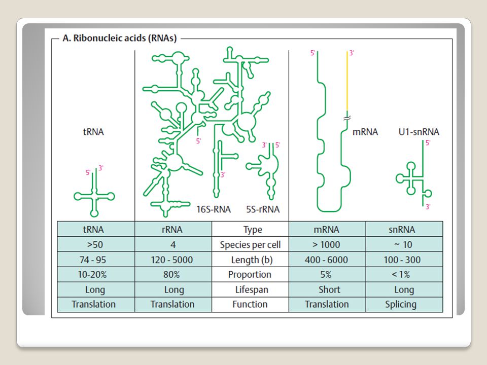
RNA Ribonucleic acids (RNAs) are polymers consisting of nucleoside phosphate components that are linked by phosphoric acid diester bonds. The bases the contain are mainly uracil, cytosine, adenine, and guanine, but many unusual and modified bases are also found in RNAs (B). RNAs do not form extended double helices. In RNAs, the base pairs usually only extend over a few residues. Large RNAs such as ribosomal 16SrRNA (center) contain numerous “stem and loop” regions of this type. These sections are again folded three- dimensionally—i. e., like proteins, RNAs have a tertiary structure
. RNAs do not form extended double helices. In RNAs, the base pairs usually only extend over a few residues. Large RNAs such as ribosomal 16SrRNA (center) contain numerous stem and loop regions of this type. These sections are again folded three- dimensionally—i. e., like proteins, RNAs have a tertiary structure.")
13
RNA Cellular RNAs vary widely in their size, structure, and lifespan. The great majority of them are ribosomal RNA (rRNA), which in several forms is a structural and functional component of ribosomes. Ribosomal RNA is produced from DNA by transcription in the nucleolus, and it is processed there and assembled with proteins to form ribosome subunits. The bacterial 16S-rRNA shown in Fig. A, with nucleotides (nt), is a component of the small ribosomae subunit, while the much smaller 5S-rRNA (118 nt) is located in the large subunit Messenger RNAs (mRNAs) transfer genetic information from the cell nucleus to the cytoplasm. The transfer RNAs (tRNAs) function during translation as links between the nucleic acids and proteins
, which in several forms is a structural and functional component of ribosomes. Ribosomal RNA is produced from DNA by transcription in the nucleolus, and it is processed there and assembled with proteins to form ribosome subunits. The bacterial 16S-rRNA shown in Fig. A, with 1542 nucleotides (nt), is a component of the small ribosomae subunit, while the much smaller 5S-rRNA (118 nt) is located in the large subunit. Messenger RNAs (mRNAs) transfer genetic information from the cell nucleus to the cytoplasm. The transfer RNAs (tRNAs) function during translation as links between the nucleic acids and proteins.")
16
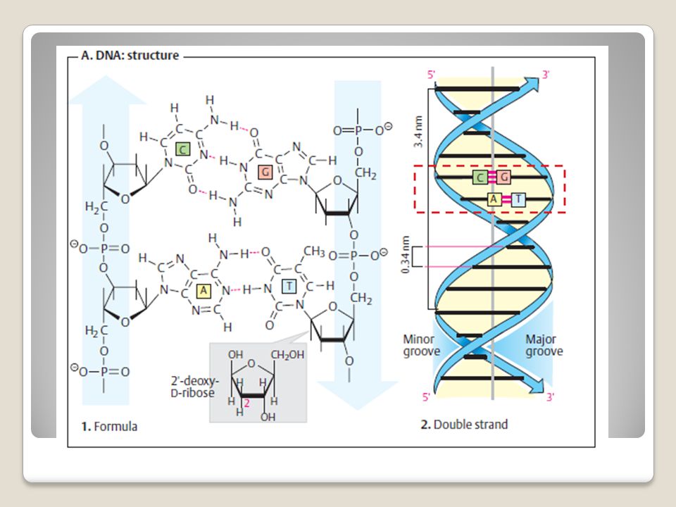
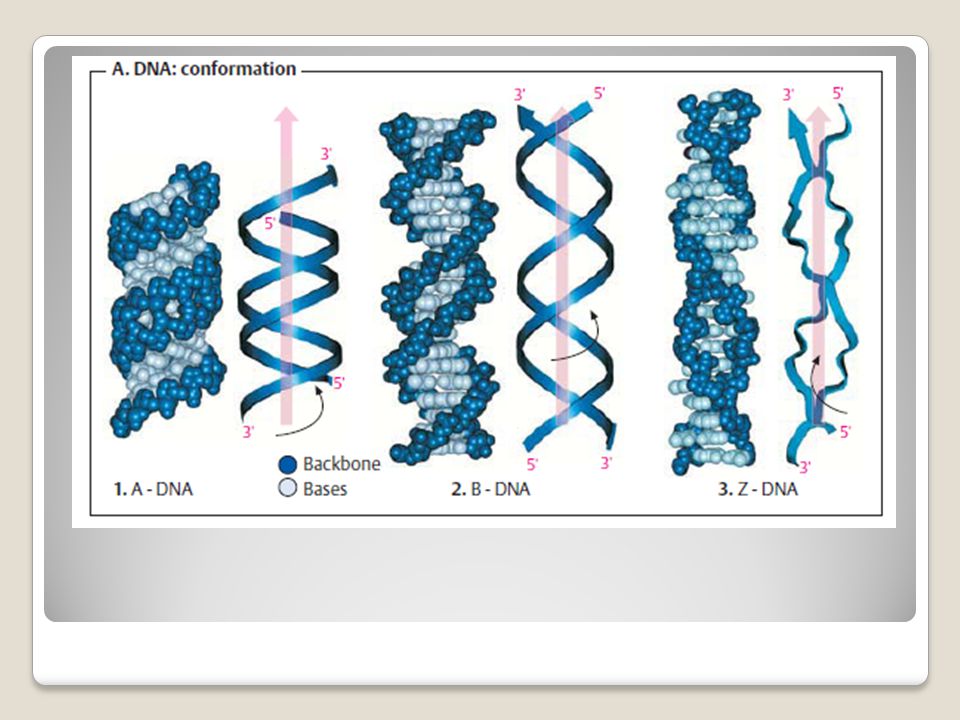
DNA (DNAs) are polymeric molecules consisting of nucleotide building blocks. DNA contains 2-deoxyribose, and the uracil base in RNA is replaced by thymine. DNA consists of two polydeoxynucleotide molecules (“strands”). Each base in one strand is linked to a comple- mentary base in the other strand by H-bond. (A =T; G= C) Potential donors are amino groups (Ade, Cyt, Gua) and ring NH groups. Possible acceptors are carbonyl oxygen atoms (Thy, Cyt, Gua) and ring nitrogen atoms. Two linear and therefore highly stable bonds can thus be formed in A–T pairs, and three in G–C pairs DNA serves to store genetic information Specific segments of DNA (“genes”) are transcribed as needed into RNAs, which either carry out structural or catalytic tasks themselves or provide the basis for synthesizing proteins
. Each base in one strand is linked to a comple- mentary base in the other strand by H-bond. (A =T; G= C) Potential donors are amino groups (Ade, Cyt, Gua) and ring NH groups. Possible acceptors are carbonyl oxygen atoms (Thy, Cyt, Gua) and ring nitrogen atoms. Two linear and therefore highly stable bonds can thus be formed in A–T pairs, and three in G–C pairs. DNA serves to store genetic information. Specific segments of DNA ( genes ) are transcribed as needed into RNAs, which either carry out structural or catalytic tasks themselves or provide the basis for synthesizing proteins.")
19
All of the words (“codons”) contain three letters (“triplets”), and each triplet stands for one of the 20 proteinogenic amino acids.
 contain three letters ( triplets ), and each triplet stands for one of the 20 proteinogenic amino acids.")
22
Degradation of nucleotides
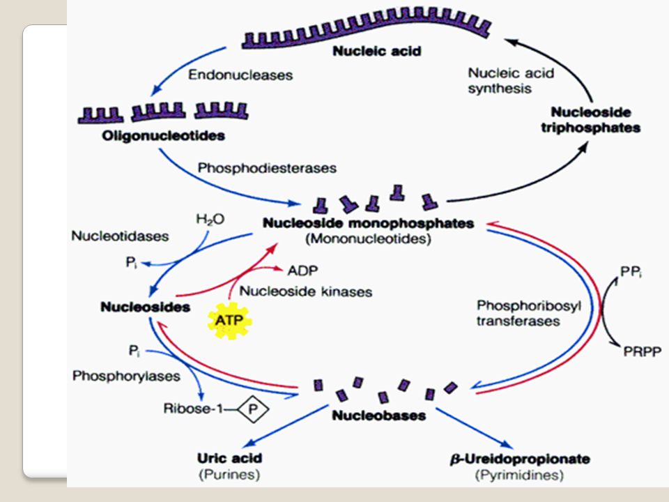
The principles underlying the degradation of purines (1) and pyrimidines (2) differ. In the human organism, purines are degraded into uric acid and excreted in this form. The purine ring remains intact in this process. In contrast, the ring of the pyrimidine bases (uracil, thymine, and cytosine) is broken down into small fragments, which can be returned to the metabolism Degradation of nucleotides
 and pyrimidines (2) differ. In the human organism, purines are degraded into uric acid and excreted in this form. The purine ring remains intact in this process. In contrast, the ring of the pyrimidine bases (uracil, thymine, and cytosine) is broken down into small fragments, which can be returned to the metabolism. Degradation of nucleotides.")
23
Purine (left). The purine nucleotide guanosine monophosphate (GMP, 1) is degraded in two steps—first to the guanosine and then to guanine (Gua). Guanine is converted by deamination into another purine base, xanthine.
 is degraded in two steps—first to the guanosine and then to guanine (Gua). Guanine is converted by deamination into another purine base, xanthine.")
24
Purine and pyrimidine biosynthesis
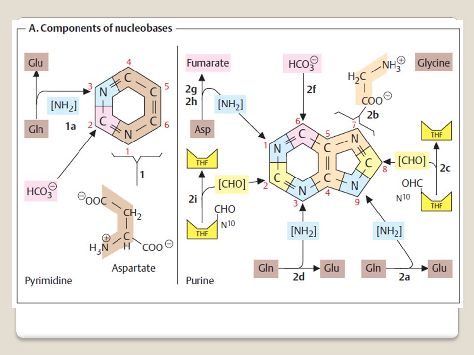
The bases occurring in nucleic acids are derivatives of the aromatic heterocyclic compounds purine and pyrimidine The major intermediates in the biosynthesis of nucleic acid components are the mononucleotides uridine monophosphate(UMP) in the pyrimidine series and inosine monophosphate (IMP, base: hypoxanthine) in the purines. The synthetic pathways for pyrimidines and purines are fundamentally different. For the pyrimidines, the pyrimidine ring is first constructed and then linked to ribose 5-phosphate to form a nucleotide. By contrast, synthesis of the purines starts directly from ribose 5- phosphate.
 in the pyrimidine series and inosine monophosphate (IMP, base: hypoxanthine) in the purines. The synthetic pathways for pyrimidines and purines are fundamentally different. For the pyrimidines, the pyrimidine ring is first constructed and then linked to ribose 5-phosphate to form a nucleotide. By contrast, synthesis of the purines starts directly from ribose 5- phosphate.")
25
Purine and pyrimidine biosynthesis
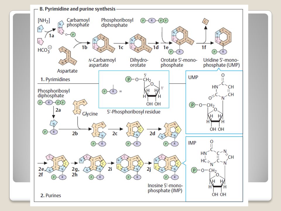
The precursors for the synthesis of the pyrimidine ring are carbamoyl phosphate, which arises from glutamate and HCO3 – (1a) and the amino acid aspartate. These two components are linked to N-carbamoyl aspartate (1b) and then converted into dihydroorotate by closure of the ring (1c). In mammals, steps 1a to 1c take place in the cytoplasm, and are catalyzed by a singlemultifunctional enzyme. In the next step (1d), dihydroorotate is oxidized to orotate by an FMN-dependent dehydrogenase. Orotate is then linked with phosphoribosyl diphosphate (PRPP) to form the nucleotide orotidine 5-monophosphate (OMP). Finally, decarboxylation yields uridine 5-monophosphate (UMP).
 and the amino acid aspartate. These two components are linked to N-carbamoyl aspartate (1b) and then converted into dihydroorotate by closure of the ring (1c). In mammals, steps 1a to 1c take place in the cytoplasm, and are catalyzed by a singlemultifunctional enzyme. In the next step (1d), dihydroorotate is oxidized to orotate by an FMN-dependent dehydrogenase. Orotate is then linked with phosphoribosyl diphosphate (PRPP) to form the nucleotide orotidine 5-monophosphate (OMP). Finally, decarboxylation yields uridine 5-monophosphate (UMP).")
26
Purine and pyrimidine biosynthesis
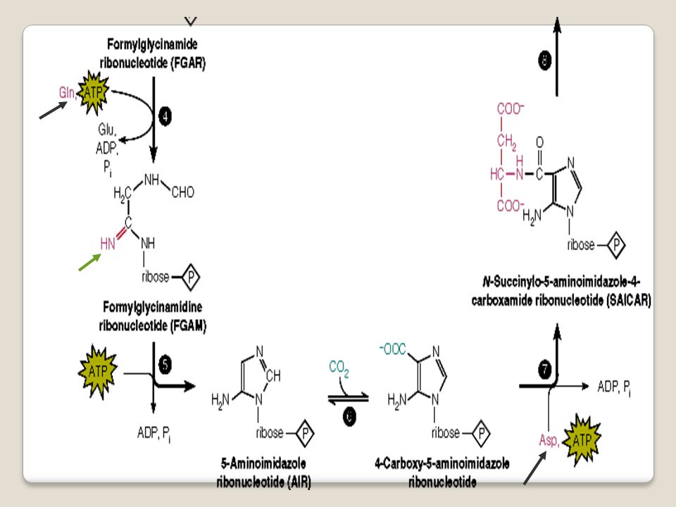
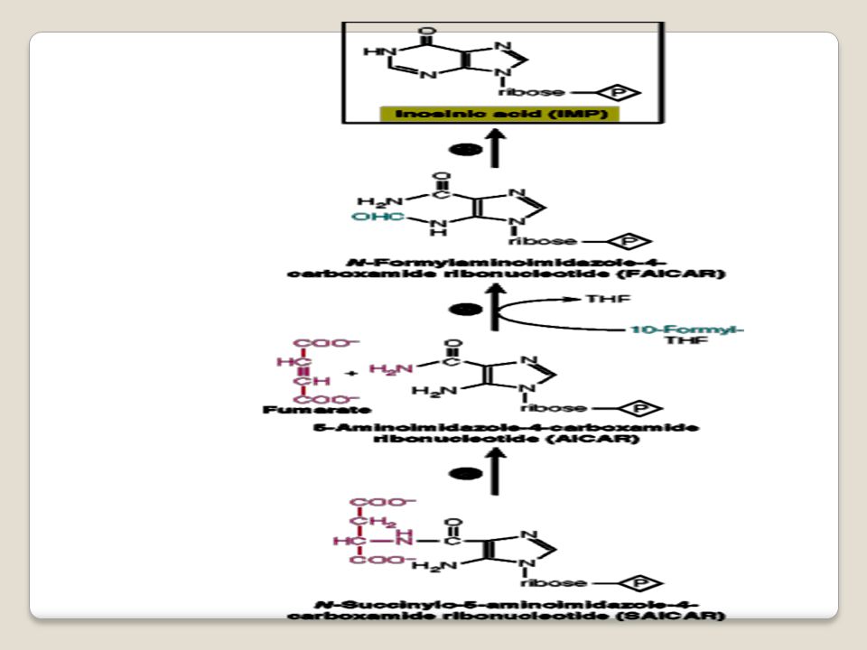
Purine biosynthesis starts with PRPP. Formation of the ring starts with transfer of an amino group, from which the later N-9 is derived (2a). Glycine and a formyl group from N10-formyl-THF then supply the remaining atoms of the five-membered ring (2b, 2c). Before the five-memberedring is closed (in step 2f), atoms N-3 and C-6 of the later six-membered ring are attached (2d, 2e). Synthesis of the ring then continues with N-1 and C-2 (2g, 2i). In the final step (2j), the six-membered ring is closed, and inosine 5-monophosphate arises. The IMP formed does not accumulate, but is rapidly converted into AMP and GMP.
. Glycine and a formyl group from N10-formyl-THF then supply the remaining atoms of the five-membered ring (2b, 2c). Before the five-memberedring is closed (in step 2f), atoms N-3 and C-6 of the later six-membered ring are attached (2d, 2e). Synthesis of the ring then continues with N-1 and C-2 (2g, 2i). In the final step (2j), the six-membered ring is closed, and inosine 5-monophosphate arises. The IMP formed does not accumulate, but is rapidly converted into AMP and GMP.")
29
Nucleotide biosynthesis
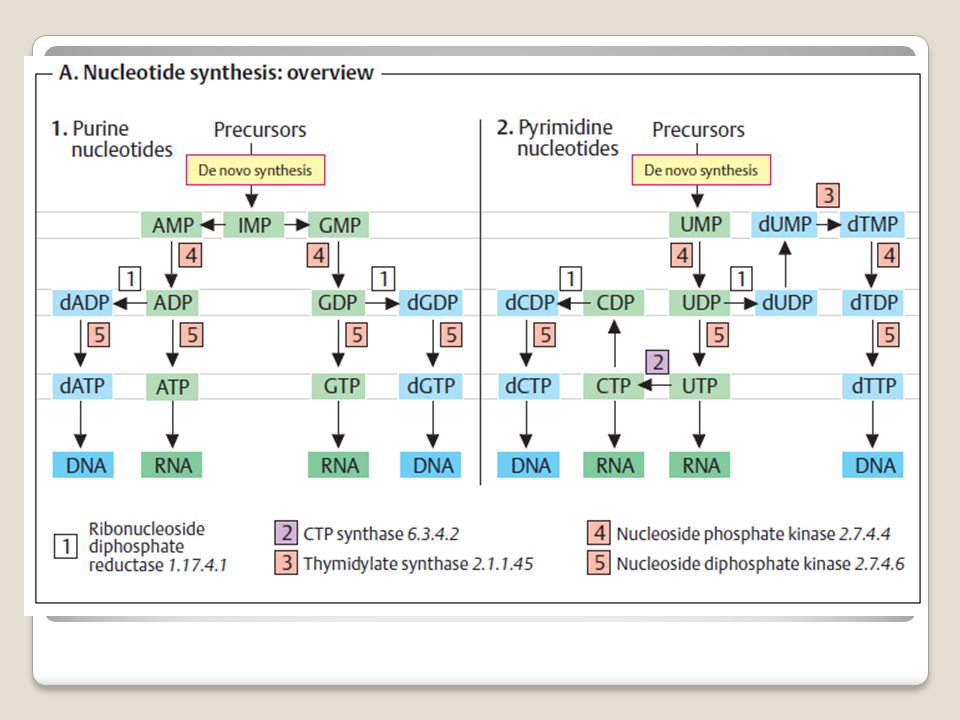
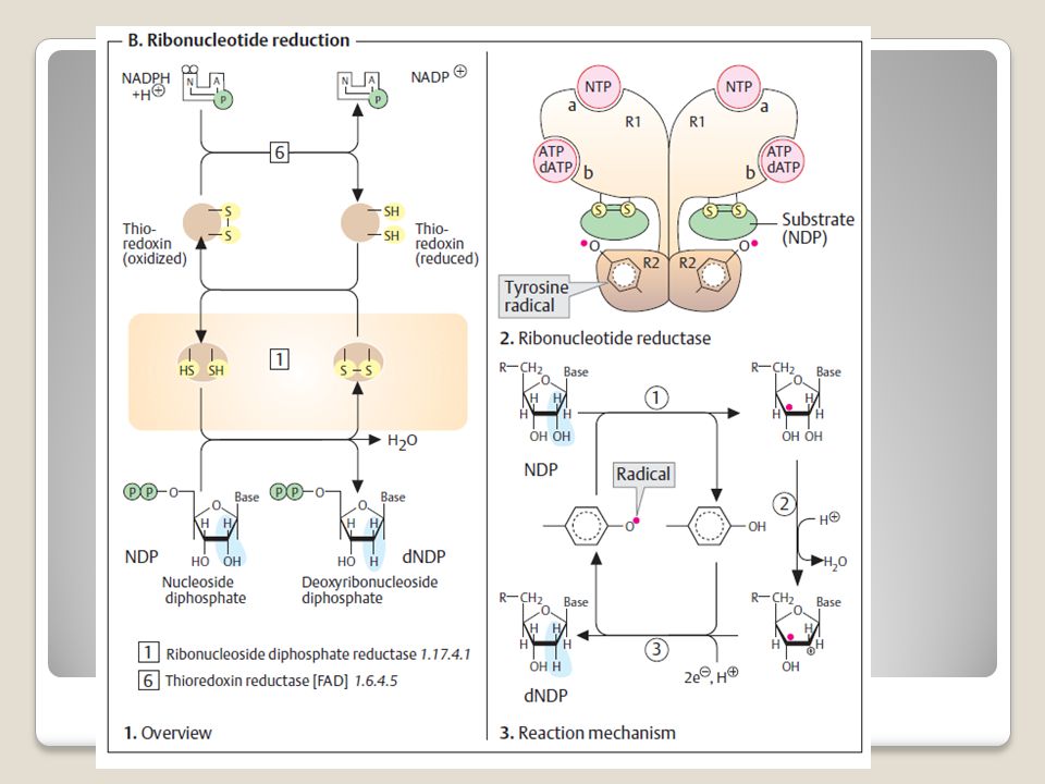
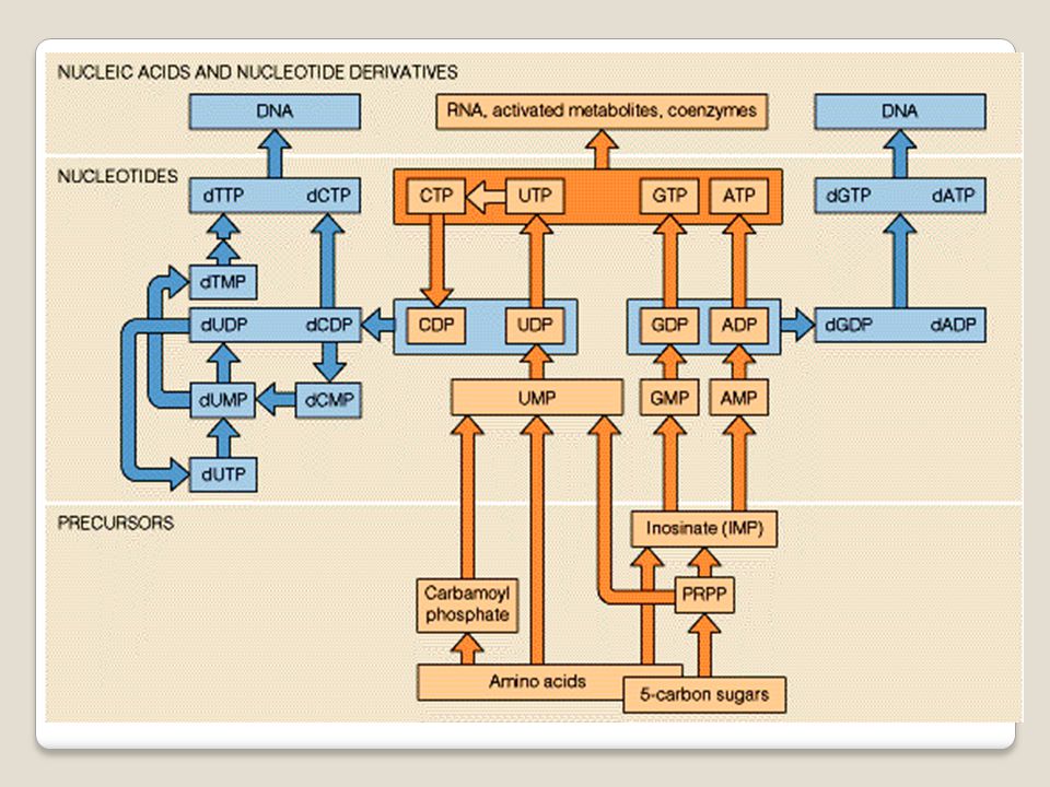
De novo synthesis of purines and pyrimidines yields the monophosphates IMP and UMP, respectively. All other nucleotides and deoxynucleotides are synthesized from these two precursors. The synthesis of purine nucleotides (1) starts from IMP. The base it contains, hypoxanthine, is converted in two steps each into adenine or guanine. The nucleoside monophosphates AMP and GMP that are formed are then phosphorylated by nucleoside phosphate kinases to yield the diphosphates ADP and GDP, and these are finally phosphorylated into the triphosphates ATP and GTP. The nucleoside triphosphates serve as components for RNA, or function as coenzymes (see p. 106). Conversion of the ribonucleotides into deoxyribonucleotides occurs at the level of the diphosphates and is catalyzed by nucleoside diphosphate reductase (B).
 starts from IMP. The base it contains, hypoxanthine, is converted in two steps each into adenine or guanine. The nucleoside monophosphates AMP and GMP that are formed are then phosphorylated by nucleoside phosphate kinases to yield the diphosphates ADP and GDP, and these are finally phosphorylated into the triphosphates ATP and GTP. The nucleoside triphosphates serve as components for RNA, or function as coenzymes (see p. 106). Conversion of the ribonucleotides into deoxyribonucleotides occurs at the level of the diphosphates and is catalyzed by nucleoside diphosphate reductase (B).")
30
The biosynthetic pathways for the pyrimidine nucleotides (2) are more complicated.
The first product, UMP, is phosphorylated first to the diphosphate and then to the triphosphate, UTP. CTP synthase then converts UTP into CTP. Since pyrimidine nucleotides are also reduced to deoxyribonucleotides at the diphosphate level, CTP first has to be hydrolyzed by a phosphatase to yield CDP before dCDP and dCTP can be produced. The DNA component deoxythymidine triphosphate (dTTP) is synthesized fromUDP in several steps. The base thymine, which only occurs in DNA, is formed by methylation of dUMP at the nucleosidemonophosphate level
 is synthesized fromUDP in several steps. The base thymine, which only occurs in DNA, is formed by methylation of dUMP at the nucleosidemonophosphate level.")
31
Ribonucleotide reduction
2-Deoxyribose, a component of DNA, is not synthesized as a free sugar, but arises at the diphosphate level by reduction of ribonucleoside diphosphates. This reduction is a complex process in which several proteins are involved. The reducing equivalents needed come from NADPH+H+. However, they are not transferred directly from the coenzyme to the substrate, but first pass through a redox series that has several steps (1). In the first step, thioredoxin reductase reduces a small redox protein, thioredoxin, via enzyme-bound FAD. This involves cleavage of a disulfide bond in thioredoxin. The resulting SH groups in turn reduce a catalytically active disulfide bond in nucleoside diphosphate reductase (“ribonucleotide reductase”). The free SH groups formed in this way are the actual electron donors for the reduction of ribonucleotide diphosphates.
. In the first step, thioredoxin reductase reduces a small redox protein, thioredoxin, via enzyme-bound FAD. This involves cleavage of a disulfide bond in thioredoxin. The resulting SH groups in turn reduce a catalytically active disulfide bond in nucleoside diphosphate reductase ( ribonucleotide reductase ). The free SH groups formed in this way are the actual electron donors for the reduction of ribonucleotide diphosphates.")
35
Metabolisme asam nukleat dan nukleotida

37
Hampir semua organisme mampu mensintesis nukleotida dr prekursor yg lebih sederhana jalur de novo untuk nukleotida mirip utk setiap organisme Nukleotida juga dapat disintesis dari hasil pemecahan nukleotida yang telah ada salvage pathway (recycle) yaitu dari degradasi pirimidin dan purin dari sel yang mati (regenerasi) atau dari makanan
 yaitu dari degradasi pirimidin dan purin dari sel yang mati (regenerasi) atau dari makanan.")
40
5-Phospho- -D-ribosyl-1-pyrophosphate (PRPP)
Intermediet untuk baik proses de novo and salvage pathway Berasal dari ribosa 5 phosphat

41
Biosintesis De Novo Purines

44
GAR synthetase IMP synthase AICAR transformylase GAR transformylase
SAICAR lyase FGAR amidotransferase SAICAR synthetase FGAM cyclase AIR karboksilase

45
Hal-hal penting dalam sintesis de novo purin:
Sangat tergantung pada “pool” ribosa Gugus amina didonor oleh glutamin dgn enzim amidotransferase Glisin dan fumarat donor ring dlm nukleotida Daur reaksi dikontrol secara alosterik dgn AMP, ADP, GMP dan GDP. bekerja pada PRPP amidotransferase

46
Daur diawali dgn perubahan PRPP IMP
IMP = Inosine monofosfat mrpkn bentuk nukleotida purin yang pertama dibentuk dlm daur ini Sebagai basa adalah hypoxanthin

47
DAUR dr IMP AMP & GMP Adenilosuksinat synthetase IMP dehidrogenase
XMP aminase Adenilosuksinat lyase DAUR dr IMP AMP & GMP

48
Metabolisme de novo nukleotida pirimidine
CP synthetase Aspartat transcarbomoylase Dihidrooratate DH dihydrorotase

49
Orotat fosforibosiltransferase
Orotidilate dekarboksilase CTP synthetase UMP kinase Nukleosida diphosphat kinase

50
Hal-hal penting dalam sintesis de novo pirimidine:
cincin pirimidine disintesis terpisah dr gula ribosa nya Daur pirimidine de novo tidak bercabang produk akhir dr daur adalah UMP yang mrpkn bahan dari CMP Reaksi pertama pembtkan karbamoyl aspartate dr asp dan carbomoyl-P titik regulasi yg penting dlm daur tsb Aspartat transcarbomoylase (ATCase) diaktivasi oleh diaktivasi oleh ATP dan dihambat oleh CTP sbg produk akhir
 diaktivasi oleh diaktivasi oleh ATP dan dihambat oleh CTP sbg produk akhir.")
51
Degradasi purine Produk akhir katabolisme purin : asam urat

52
Degradasi pirimidin

54
Metabolisme asam nukleat II

55
Merupakan proses metabolisme informasi, yang berbeda dgn metabolisme- metabolisme yang telah dipelajari sebelumnya: metabolisme intermediate ensim berperanan dlm setiap reaksi yg terjadi. Proses perlekatan substrat dan menghasilkan produk Metabolisme informasi ada cetakan yang perlu diterjemahkan menjadi produk. Cetakan DNA atau RNA, proses juga melibatkan berbagai enzim

56
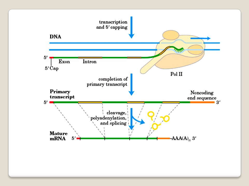
Proses utama dlm metabolisme informasi:
Replikasi DNA berperan sbg cetakan untuk sintesisnya sdr Transkripsi Informasi yang ada pada DNA menentukan RNA yang diproduksi Translasi RNA berperan sbg cetakan untuk sintesis suatu rantai polipeptida ttt

57
Replikasi dan transkripsi hanya menggunakan 4 nukleotida
Translasi mengubah bahasa nukleotida yg terdiri dari 4 nukleotida menjadi bahasa protein yang terdiri dari 20 huruf asam amino Persamaan replikasi, transkripsi dan translasi membutuhkan cetakan proses terdiri dari inisiasi, elongasi dan terminasi

58
Replikasi Secara konsep sederhana Proses mekanismenya komplek
Kesederhanaannya krn konsep dr Watson & Crick Transfer informasi melibatkan pembukaan double helix DNA yang diikuti secara bersamaan dengan pembentukan dua pita baru pasangan dari pita DNA yang lama Replikasi

59
Replikasi dimulai pada suatu lokasi tertentu
arah dari replikasi tidak semuanya sama Sintesis DNA selalu dengan arah 5’ 3” leading strand disintesis secara kontinyu lagging strand disintesis secara diskontinyu okzaki fragment

60
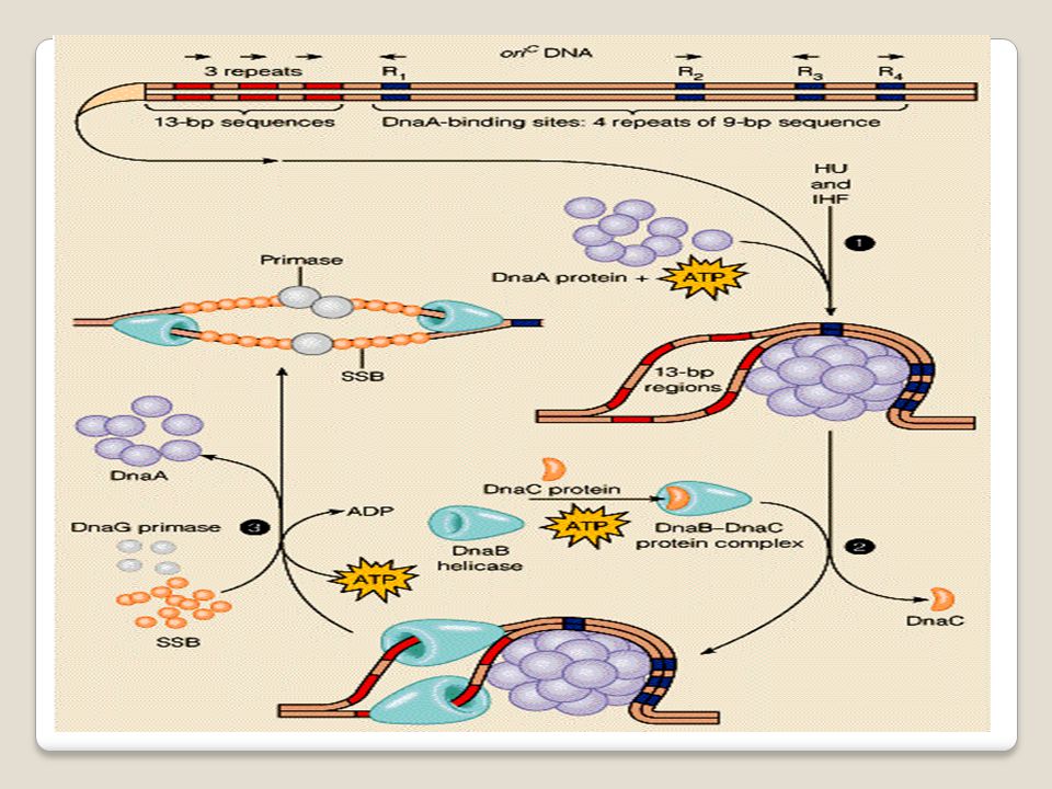
Proses inisiasi replikasi DNA
Urutan nukleotida yang secara spesifik terikat pada protein inisiasi Mekanisme untuk mensintesi primer RNA dpt dielongasi oleh DNA polimerase Inisiasi DNA replikasi pada E coli Ori C Proses inisiasi replikasi DNA

65
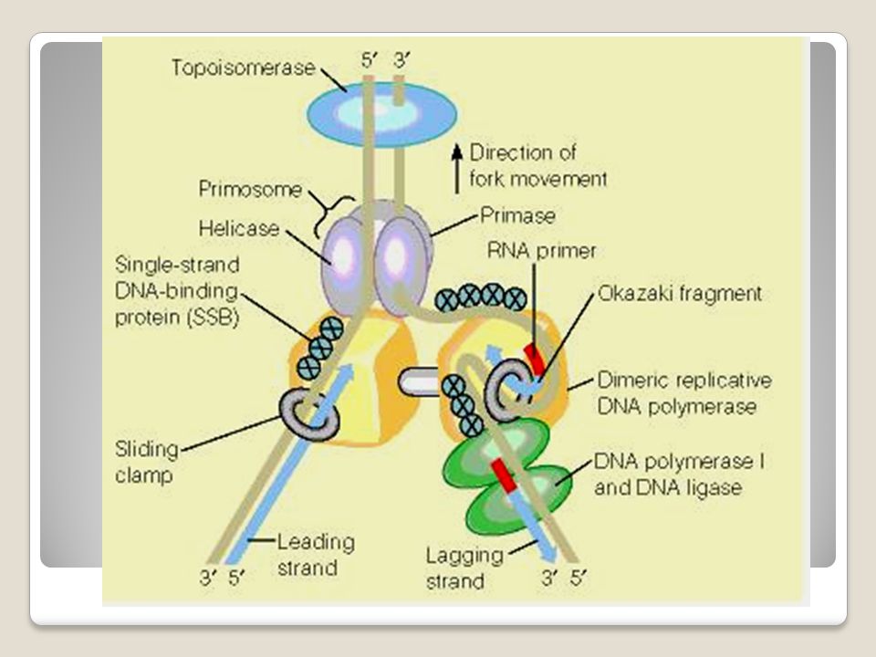
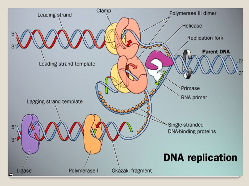
Helicase membuka double helix DNA
Primase mensintesis primer RNA Topoisomerase melepaskan torsi krn proses membukanya DNA DNA polymerase dimer, melakukan elongasi baik pd lagging dan leading strand Sliding clamp memegang rantai polipeptida baru dengan cetakannya Single strand DNA binding Protein SSBP menstabilkan cetakan DNA memfasilitasi pengikatan nukleotida baru

66
DNA polimerasi I menghilangkan RNA primer yang melekat pada lagging strand DNA dan mengganti dgn DNA, DNA Ligase menyambung DNA antara okazaki fragment satu dgn yg lain

68
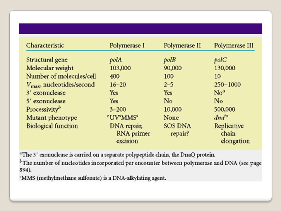
DNA polimerase Pada sel bakteri dikenal ada 3 macam DNA polimerase
DNA polimerase I, II dan III DNA polimerase I mempunyai aktivitas eksonuklease proof reading

70
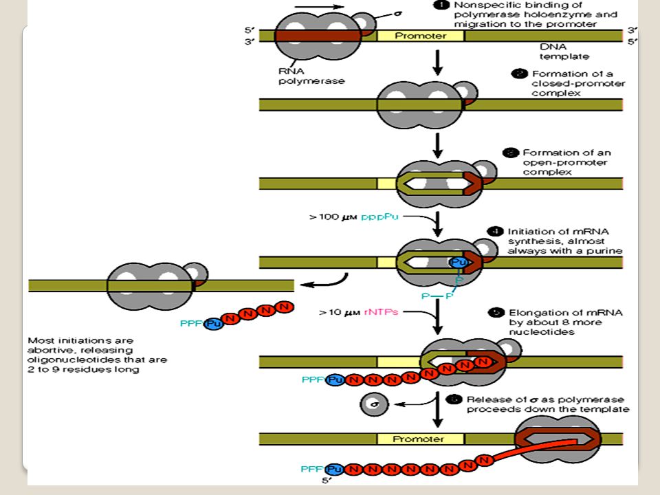
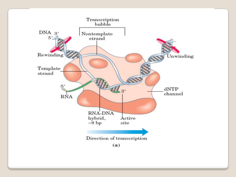
Suatu proses untuk membaca informasi yang disimpan dalam urutan nukleotida DNA RNA
RNA sintesis membutuhkan ensim RNA polimerase Mekanisme dibagi menjadi 3 Inisiasi Elongasi Terminasi Transkripsi DNA

77
Translation adalah proses membaca kodon dan menggabungkan asam amino yang sesuai bersama-sama dengan ikatan peptida. Komponen proses translasi mRNA consist of genetic code Ribosome tRNA together with a.a Enzymes Translasi DNA

78
Elongation Termination Translation process consists of 3 main stages
Initiation Elongation Termination Initiation Activation of amino acids for incorporation into proteins.

79
Activation of amino acids for incorporation into proteins.

80
Genetic code 3 nucleotides - codon – mengkode untuk 1 asam amino dlm suatu protein
Codon urutan 3 nukleotida dalam mRNA yang menspesifikasikan penggabungan suata asam amino ttt mjd protein. The relationship between codons and the amino acids they code for is called the genetic code.

81
Not all codons are used with equal frequency.
There is a considerable amount of variation in the patterns of codon usage between different organisms.

82
Relationships of DNA to mRNA to polypeptide chain.

83
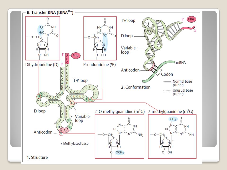
Translation is accomplished by the anticodon loop of tRNA forming base pairs with the codon of mRNA in ribosomes

84
Transfer RNA (tRNA) composed of a nucleic acid and
a specific amino acid provide the link between the nucleic acid sequence of mRNA and the amino acid sequence it codes for. An anticodon a sequence of 3 nucleotides in a tRNA that is complementary to a codon of mRNA Structure of tRNAs

85
Only tRNAfMet is accepted to form the initiation complex.
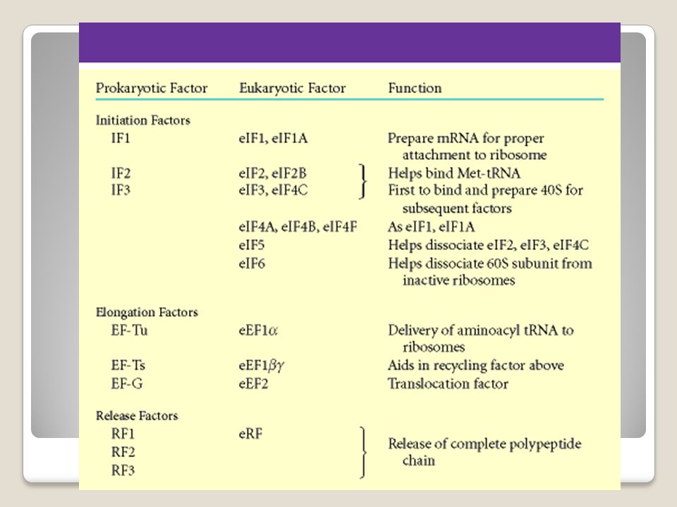
All further charged tRNAs require fully assembled (i.e., 70S) ribosomes The Shine-Dalgarno sequence help ribosomes and mRNA aligns correctly for the start of translation. Ribosome consists of A site aminoacyl P site peptidyl - E site exit Two initiation factors (IF1 &IF3) bind to a 70S ribosome. promote the dissociation of 70S ribosomes into free 30S and 50S subunits. mRNA and IF2, which carries GTP the charged tRNA bind to a free 30S subunit. After these have all bound, the 30S initiation complex is complete.
 ribosomes. The Shine-Dalgarno sequence help ribosomes and mRNA aligns correctly for the start of translation. Ribosome consists of. A site aminoacyl. P site peptidyl. - E site exit. Two initiation factors (IF1 &IF3) bind to a 70S ribosome. promote the dissociation of 70S ribosomes into free 30S and 50S subunits. mRNA and IF2, which carries. GTP. the charged tRNA. bind to a free 30S subunit. After these have all bound, the 30S initiation complex is complete.")
86
Peptide bond formation
catalyzed by an enzyme complex called peptidyltransferase Peptidyltransferase consists of some ribosomal proteins and the ribosomal RNA acts as a ribozyme. The process is repeated until a termination signal is reached.

87
Termination of translation occurs when one of the stop codons (UAA, UAG, or UGA) appears in the A site of the ribosome. No tRNAs correspond to those sequences, so no tRNA is bound during termination. Proteins called release factors participate in termination

89
Terima kasih

Presentasi serupa