Upload presentasi
Presentasi sedang didownload. Silahkan tunggu

1
FISIOLOGI SISTEM INTEGUMEN
dr. Nindya Aryanty, M.Med.Ed

2
Pokok Bahasan Gambaran umum ttg sistem integumen Epidermis Dermis
Lapisan subkutan Struktur asesorius

3
A. Gambaran Umum Tentang Sistem Integumen

4
Tujuan pembelajaran- Mahasiswa mampu:
Menjelaskan komponen-komponen dalam sistem integumen Menjelaskan dengan rinci fungsi sistem integumen

5
Sistem integumen adalah sistem organ yang paling aksessibel, luas, dan memiliki fungsi yang bervariasi 16% total BB, luas 1,5-2 meter persegi Terdiri dari 2 komponen utama: a. Membrana kutaneus: - epidermis - dermis b. Struktur asesorius: - rambut - kuku - kelenjar eksokrin

6
Komponen-komponen sistem integumen

7
Di bawah lapisan dermis jaringan ikat longgar = lapisan subkutan (hypodermis)
Hypodermis memisahkan integumen dari fascia otot atau tulang

8
Fungsi kulit dan lapisan subkutan:
Memproteksi jaringan & organ di bawahnya dari benturan, perlukaan, kehilangan cairan, trauma kimia Ekskresi garam, air, organic waste oleh kelenjar integumen Mempertahankan suhu tubuh normal Sintesis vitamin D utk pembentukan hormon calcitriol (metabolisme kalsium) Penyimpanan lemak di dalam adipocytes dari dermis dan lapisan subkutan Mendeteksi sentuhan, tekanan, nyeri, stimuli suhu disampaikan pada sistem saraf
 Penyimpanan lemak di dalam adipocytes dari dermis dan lapisan subkutan. Mendeteksi sentuhan, tekanan, nyeri, stimuli suhu disampaikan pada sistem saraf.")
9
B. Epidermis

10
Tujuan Pembelajaran- Mahasiswa mampu:
Menjelaskan struktur utama dari epidermis dan fungsi masing-masing lapisannya Menjelaskan mekanisme pewarnaan kulit Menjelaskan efek sinar UV pada kulit, dan menjelaskan peran melanosit saat kulit terpapar sinar matahari

11
Epidermis terdiri dari EPITEL SKUAMOUS BERLAPIS (stratified squamous epithelium)
Provides mechanical protection and also helps keep microorganisms outside the body Avaskular tergantung pada proses difusi nutrien & oksigen dari kapiler-kapiler di dalam dermis The epidermal cells with the highest metabolic demands are found close to the basement membrane, where the diffusion distance is short. The superficial cells, far removed from the source of nutrients, are dead.

12
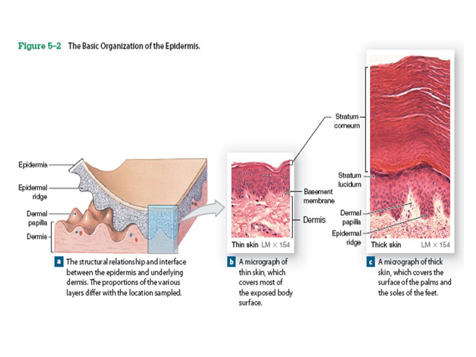
Strata: - stratum basale, - stratum spinosum - stratum granulosum
Epidermis didominasi oleh keratinosit (sel mengandung protein keratin) membentuk lapisan-lapisan (strata) Strata: - stratum basale, - stratum spinosum - stratum granulosum - stratum lucidum - stratum corneum. Berdasarkan ketebalan epidermis, Kulit dibagi menjadi: Thin skin 4 lapisan keratinosit, 0,08 mm Thick skin lapisan ke-5 (lapisan lusidum), 0,5 mm
 membentuk lapisan-lapisan (strata) Strata: - stratum basale, - stratum spinosum. - stratum granulosum. - stratum lucidum. - stratum corneum. Berdasarkan ketebalan epidermis, Kulit dibagi menjadi: Thin skin 4 lapisan keratinosit, 0,08 mm. Thick skin lapisan ke-5 (lapisan lusidum), 0,5 mm.")
14
B. Epidermis: 1. Stratum basale (Stratum germinativum) 2
B. Epidermis: 1. Stratum basale (Stratum germinativum) 2. stratum spinosum 3. Stratum granulosum 4. Stratum lucidum 5. Stratum corneum
 2. stratum spinosum 3. Stratum granulosum 4. Stratum lucidum 5. Stratum corneum.")
15
The stratum basale and the underlying dermis interlock, increasing the strength of the bond between the epidermis and dermis. The stratum basale forms epidermal ridges, which extend into the dermis and are adjacent to dermal projections called dermal papillae (singular, papilla; a nipple-shaped mound).
.")
16
The contours of the skin surface follow the ridge patterns, which vary from small pattern (in thin skin) to the complex whorls seen on the thick skin of the palms and soles. Ridges on the palms and soles increase the surface area of the skin and increase friction, ensuring a secure grip. Ridge shapes are genetically determined. The pattern of one epidermal ridges is unique and does not change during lifetime.

17
Sel Basal (sel germinativum)
mendominasi stratum basale. merupakan stem cells yang tumbuh (berdivisi) dan dapat naik untuk menggantikan keratinosit yang terletak lebih superfisial yang hilang atau rusak pada permukaan epitelial. Sel Merkel (sel taktil) Permukaan kulit yang tidak banyak terdapat rambut juga mengandung sel eptelial khusus tactile cells (Merkel cells) yang tersebar di antara sel-sel lainnya di stratum basale. Tactile cells are sensitive to touch; when compressed, they release chemicals that stimulate sensory nerve endings. Melanosit Pewarnaan coklat pada kulit berasal dari aktifitas sintetik dari sel pigmen yang disebut melanosit (melanocytes) yang terdistribusi di seluruh stratum basale yang dapat bergerak perlahan ke lapisan yang lebih superfisial
 dan dapat naik untuk menggantikan keratinosit yang terletak lebih superfisial yang hilang atau rusak pada permukaan epitelial. Sel Merkel (sel taktil) Permukaan kulit yang tidak banyak terdapat rambut juga mengandung sel eptelial khusus tactile cells (Merkel cells) yang tersebar di antara sel-sel lainnya di stratum basale. Tactile cells are sensitive to touch; when compressed, they release chemicals that stimulate sensory nerve endings. Melanosit. Pewarnaan coklat pada kulit berasal dari aktifitas sintetik dari sel pigmen yang disebut melanosit (melanocytes) yang terdistribusi di seluruh stratum basale yang dapat bergerak perlahan ke lapisan yang lebih superfisial.")
18
B. Epidermis: 1. Stratum basale (Stratum germinativum) 2
B. Epidermis: 1. Stratum basale (Stratum germinativum) 2. stratum spinosum 3. Stratum granulosum 4. Stratum lucidum 5. Stratum corneum
 2. stratum spinosum 3. Stratum granulosum 4. Stratum lucidum 5. Stratum corneum.")
19
Each time a stem cell divides, one of the daughter cells is pushed superficial to the stratum basale into the stratum spinosum which consists of 8 to 10 layers of keratinocytes Some of the cells entering this layer from the stratum basale continue to divide, further increasing the thickness of the epithelium. The stratum spinosum also contains dendritic (Langerhans) cells, which participate in the immune response by stimulating a defense against (1) microorganisms that manage to penetrate the superficial layers of the epidermis and (2) superficial skin cancers
 cells, which participate in the immune response by stimulating a defense against. (1) microorganisms that manage to penetrate the superficial layers of the epidermis and. (2) superficial skin cancers.")
20
B. Epidermis: 1. Stratum basale (Stratum germinativum) 2
B. Epidermis: 1. Stratum basale (Stratum germinativum) 2. stratum spinosum 3. Stratum granulosum 4. Stratum lucidum 5. Stratum corneum
 2. stratum spinosum 3. Stratum granulosum 4. Stratum lucidum 5. Stratum corneum.")
21
Stratum granulosum terdiri dari 3-5 lapisan keratinosit yang berasal dari stratum spinosum.
Ketika sel mencapai lapisan ini, sel berhenti membelah diri dan mulai memproduksi sejumlah besar protein keratin (keros, horn) & keratohyalin . Keratin, merupakan protein yang keras dan fibrous yang merupakan komponen dari struktur dasar rambut & kuku Ketika serat-serat keratin mulai dibentuk, sel menjadi lebih tipis dan pipih, sedangkan membrannya menjadi tebal dan menjadi kurang permeabel. Keratohyalin membentuk granula sitoplasma yang padat yang menyebabkan terjadinya dehidrasi di dalam sel sehingga mendorong agregasi dan terhubungnya antar serat keratin. Inti sel & organela lain dalam sel mati Proses dehidrasi yang terus berlanjut menyebabkan lapisan sel semakin menyatu & ‘terkunci’ satu sama lain dengan dikelilingi oleh keratohyalin.
 & keratohyalin . Keratin, merupakan protein yang keras dan fibrous yang merupakan komponen dari struktur dasar rambut & kuku. Ketika serat-serat keratin mulai dibentuk, sel menjadi lebih tipis dan pipih, sedangkan membrannya menjadi tebal dan menjadi kurang permeabel. Keratohyalin membentuk granula sitoplasma yang padat yang menyebabkan terjadinya dehidrasi di dalam sel sehingga mendorong agregasi dan terhubungnya antar serat keratin. Inti sel & organela lain dalam sel mati. Proses dehidrasi yang terus berlanjut menyebabkan lapisan sel semakin menyatu & ‘terkunci’ satu sama lain dengan dikelilingi oleh keratohyalin.")
22
B. Epidermis: 1. Stratum basale (Stratum germinativum) 2
B. Epidermis: 1. Stratum basale (Stratum germinativum) 2. stratum spinosum 3. Stratum granulosum 4. Stratum lucidum 5. Stratum corneum
 2. stratum spinosum 3. Stratum granulosum 4. Stratum lucidum 5. Stratum corneum.")
23
In the thick skin of the palms and soles, a glassy stratum lucidum (“clear layer”) covers the stratum granulosum. The cells in the stratum lucidum are flattened, densely packed, largely devoid of organelles, and filled with keratin.

24
B. Epidermis: 1. Stratum basale (Stratum germinativum) 2
B. Epidermis: 1. Stratum basale (Stratum germinativum) 2. stratum spinosum 3. Stratum granulosum 4. Stratum lucidum 5. Stratum corneum
 2. stratum spinosum 3. Stratum granulosum 4. Stratum lucidum 5. Stratum corneum.")
25
At the exposed surface of both thick skin and thin skin is the stratum corneum( cornu, horn).
It normally contains 15 to 30 layers of keratinized cells. Keratinization, or cornification, is the formation of protective, superficial layers of cells filled with keratin. This process occurs on all exposed skin surfaces except the anterior surfaces of the eyes. It takes 7 to 10 days for a cell to move from the stratum basale to the stratum corneum. The dead cells generally remain in the exposed stratum corneum for an additional two weeks before they are shed or washed away

26
Normally, the surface of the stratum corneum is relatively dry, so it is unsuitable for the growth of many microorganisms. Maintenance of this barrier involves coating the surface with lipid secretions from sebaceous glands. The stratum corneum is water resistant, but not waterproof. Water from interstitial fluids slowly penetrates to the surface, to be evaporated into the surrounding air. (insensible perspiration) 500 mL each day. In contrast, you are usually very aware of the sensible perspiration produced by active sweat glands.
 500 mL each day. In contrast, you are usually very aware of the sensible perspiration produced by active sweat glands.")
27
When the skin is immersed in water, osmotic forces may move water into or out of the epithelium. Sitting in a freshwater bath causes water to move into the epidermis, because fresh water is hypotonic (has fewer dissolved materials) compared with body fluids. The epithelial cells of the stratum corneum may swell to 4x their normal volumes noticeable in the thickly keratinized areas of the palms and soles. Swimming in the ocean reverses the direction of osmotic flow; because the ocean is a hypertonic solution, water leaves the body, crossing the epidermis from the underlying tissues. The process is slow, but long-term exposure to seawater endangers survivors of a shipwreck by accelerating dehydration.

28
B. Epidermis: Pewarnaan Kulit

29
The epidermis contains variable quantities of two pigments: carotene and melanin.
Carotene is an orange-yellow pigment that normally accumulates in epidermal cells. most apparent in cells of the stratum corneum of light-skinned individuals, but it also accumulates in fatty tissues in the deep dermis and subcutaneous layer. Carotene is found in a variety of orange vegetables, such as carrots and squashes, and thus the skin of individuals who eat lots of carrots can actually turn orange from an overabundance of carotene. The color change is very striking in pale-skinned individuals, but less obvious in people with darker skin pigmentation. Carotene can be converted to vitamin A, which is required for both the normal maintenance of epithelia and the synthesis of photoreceptor pigments in the eye.

30
Melanin is a brown, yellow-brown, or black pigment produced by melanocytes located in the stratum basale.

31
Melanocytes manufacture melanin and package it in intracellular vesicles called melanosomes.
These vesicles travel within the processes of melanocytes and are transferred intact to keratinocytes. The transfer of pigmentation colors the keratinocyte temporarily, until the melanosomes are destroyed by fusion with lysosomes.

32
In individuals with pale skin, this transfer occurs in the stratum basale and stratum spinosum, and the cells of more superficial layers lose their pigmentation. In dark-skinned people, the melanosomes are larger and the transfer may occur in the stratum granulosum as well; skin pigmentation is thus darker and more persistent.

33
The ratio of melanocytes to basal cells ranges between 1:4 and 1:20, depending on the region of the body. The skin covering most areas of the body has about 1000 melanocytes per square millimeter. The cheeks and forehead, the nipples, and the genital region (the scrotum of males and the labia majora of females) have higher concentrations (about 2000 per square millimeter). The differences in skin pigmentation among individuals do not reflect different numbers of melanocytes, but merely different levels of synthetic activity. Even the melanocytes of albino individuals are distributed normally, although the cells are incapable of producing melanin.
 have higher concentrations (about 2000 per square millimeter). The differences in skin pigmentation among individuals do not reflect different numbers of melanocytes, but merely different levels of synthetic activity. Even the melanocytes of albino individuals are distributed normally, although the cells are incapable of producing melanin.")
34
The melanin in keratinocytes protects your epidermis and dermis from the harmful effects of sunlight, which contains significant amounts of ultraviolet (UV) radiation. A small amount of UV radiation is beneficial, because it stimulates the epidermal production of a compound required for calcium ion homeostasis. However, UV radiation can also damage DNA, causing mutations and promoting the development of cancer. Within keratinocytes, melanosomes become concentrated in the region around the nucleus, where the melanin pigments act like a sunshade to provide some UV protection for the DNA in those cells. Dark-skinned individuals have greater initial protection against the effects of UV radiation

35
Over time, cumulative damage to the integument by UV exposure can harm fibroblasts, causing impaired maintenance of the dermis. The resulting structural alterations lead to premature wrinkling. In addition, skin cancers can develop from chromosomal damage in basal cells or melanocytes. One of the major consequences of the global depletion of the ozone layer increase in the rates of skin cancers (such as malignant melanoma). limiting UV exposure through a combination of protective clothing and sunscreens (or, better yet, sunblocks) is recommended during outdoor activities
. limiting UV exposure through a combination of protective clothing and sunscreens (or, better yet, sunblocks) is recommended during outdoor activities.")
36
Although too much sunlight can damage epithelial cells and deeper tissues, limited exposure to sunlight is beneficial. When exposed to ultraviolet radiation, epidermal cells in the stratum spinosum and stratum basale convert a cholesterol-related steroid into cholecalciferol , or vitamin D3. The liver then converts cholecalciferol into an intermediary product used by the kidneys to synthesize the hormone calcitriol Calcitriol is essential for the normal absorption of calcium and phosphorus by the small intestine; an inadequate supply leads to impaired bone maintenance and growth.

37
Rickets. a disease caused by vitamin D3 deficiency, results in the bending of abnormally weak and flexible bones under the weight of the body, plus other structural changes

38
C. Dermis

39
The dermis has two major components:
The papillary layer, Consists of areolar tissue, contains the capillaries, lymphatics, and sensory neurons that supply the surface of the skin. The reticular layer Consists of an interwoven meshwork of dense irregular connective tissue containing both collagen and elastic fibers. The presence of collagen and elastic fibers give the dermis strength and elasticity. The elastic fibers provide flexibility, and the collagen fibers limit that flexibility to prevent damage to the tissue Aging, hormones, and the destructive effects of UV radiation permanently reduce the amount of elastin in the dermis wrinkles & sagging skin The extensive distortion of the dermis that occurs over the abdomen during pregnancy or after substantial weight gain can exceed the elastic limits of the skin prevents it from recoiling to its original size after delivery or weight loss stretch marks

40
Accessory organs of epidermal origin, such as hair follicles and sweat glands, extend into the dermis. In addition, the reticular and papillary layers of the dermis contain networks of blood vessels and nerve fibers. Because of the abundance of sensory receptors in the skin, regional infection or inflammation can be very painful.

41
The Dermal Blood Supply
Arteries supplying the skin form networks in the hypodermis along its border with the reticular layer of the dermis called the cutaneous plexus Tributaries of these arteries supply both the adipose tissues of the subcutaneous layer and the tissues of the integument. As small arteries travel toward the epidermis, branches supply the hair follicles, sweat glands, and other structures in the dermis Trauma to the skin often results in a contusion, or bruise (“black and blue” color) result of the rupture of dermal blood vessels
 result of the rupture of dermal blood vessels.")
42
Innervation of the Skin
The integument is filled with sensory receptors, and anything that comes in contact with the skin initiates a nerve impulse that can reach our conscious awareness. Nerve fibers in the skin control blood flow adjust gland secretion rates monitor sensory receptors in the dermis and the deeper layers of the epidermis

43
Innervation of the Skin
Sensory neuron in epidermis : tactile cells (with sensory terminals known as tactile discs monitor these cells); sensory neurons that provide sensations of pain and temperature. Sensory neuron in dermis : Receptors sensitive to light touch—tactile corpuscles, located in dermal papillae— Receptors sensitive to deep pressure and vibration—lamellated corpuscles, in the reticular layer
; sensory neurons that provide sensations of pain and temperature. Sensory neuron in dermis : Receptors sensitive to light touch—tactile corpuscles, located in dermal papillae— Receptors sensitive to deep pressure and vibration—lamellated corpuscles, in the reticular layer.")
44
D. Hypodermis

45
The connective tissue fibers of the reticular layer are extensively interwoven with those of the hypodermis. Stabilizing the position of the skin in relation to underlying tissues, such as skeletal muscles or other organs The hypodermis consists of areolar and adipose tissues quite elastic. Only its superficial region contains large arteries and veins. The venous circulation of this region contains a substantial amount of blood blood reservoir The rest of the hypodermis contains a limited number of capillaries and no vital organs subcutaneous injection Subcutaneous fat also serves as a substantial energy reserve and as a shock absorber

46
E. Accessory Structures Of The Integument
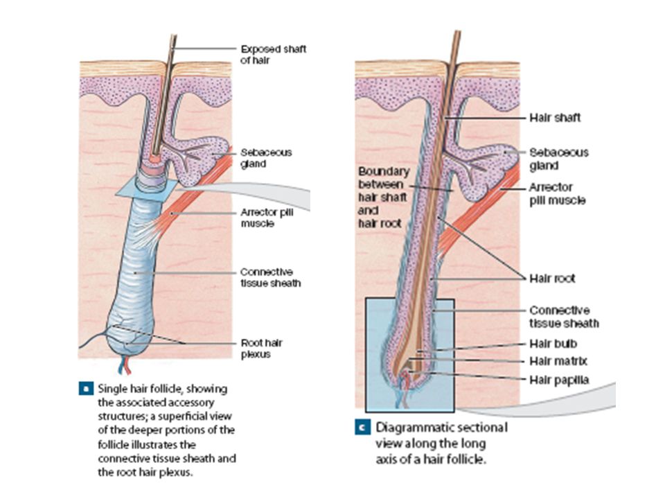
Hair—hair follicles, Sebaceous glands Sweat glands Nails located in the dermis, project through the epidermis to the integumentary surface

47
Hairs Hairs project above the surface of the skin almost everywhere, except over the sides and soles of the feet, the palms of the hands, the sides of the fingers and toes, the lips, and portions of the external genitalia. The human body has about 2.5 million hairs, and 75 percent of them are on the general body surface, not on the head. Hairs are nonliving structures produced in organs called hair follicles.

48
Hairs-function Protect scalp from ultraviolet radiation,
help cushion light impacts to the head insulate the skull The hairs guarding the entrances to nostrils and external ear canals help prevent the entry of foreign particles and insects, and also eyelashes Eyebrows help keep sweat out of eyes. As sensory receptors A root hair plexus of sensory nerves surrounds the base of each hair follicle When stimulated, the arrector pili muscle contracts, pulling on the follicle and forcing the hair to stand erect the result of emotional states, such as fear or rage, or a response to cold, producing “goose bumps.”

50
Sebaceous Glands Sebaceous glands, or oil glands,
discharge an oily lipid secretion (sebum) into hair follicles Sebaceous glands that communicate with a single follicle share a duct simple branched alveolar glands Sebum inhibits the growth of bacteria, lubricates and protects the keratin of the hair shaft, and conditions the surrounding skin Sebaceous follicles are large sebaceous glands that are not associated with hair follicles; their ducts discharge sebum directly onto the epidermis located on the face, back, chest, nipples, and external genitalia.
 into hair follicles. Sebaceous glands that communicate with a single follicle share a duct simple branched alveolar glands. Sebum inhibits the growth of bacteria, lubricates and protects the keratin of the hair shaft, and conditions the surrounding skin. Sebaceous follicles are large sebaceous glands that are not associated with hair follicles; their ducts discharge sebum directly onto the epidermis located on the face, back, chest, nipples, and external genitalia.")
51
Sebaceous Glands

52
Sweat Glands/Sudoriferous Glands
Two types of sweat glands: 1. Apocrine Sweat Glands In the armpits (axillae), around the nipples, and in the pubic region, apocrine sweat glands secrete their products (a sticky, cloudy, and potentially odorous secretion) into hair follicles Apocrine sweat glands begin secreting at puberty. The sweat produced is a nutrient source for bacteria intensify its odor. 2. Merocrine Sweat Glands (eccrine sweat glands) Discharge their secretions directly onto the surface of the skin Are far more numerous and widely distributed than apocrine sweat glands The palms and soles have the highest numbers
, around the nipples, and in the pubic region, apocrine sweat glands secrete their products (a sticky, cloudy, and potentially odorous secretion) into hair follicles. Apocrine sweat glands begin secreting at puberty. The sweat produced is a nutrient source for bacteria intensify its odor. 2. Merocrine Sweat Glands (eccrine sweat glands) Discharge their secretions directly onto the surface of the skin. Are far more numerous and widely distributed than apocrine sweat glands. The palms and soles have the highest numbers.")
53
Sweat Glands/Sudoriferous Glands
The functions of merocrine sweat gland : • Cooling the Surface of the Skin to Reduce Body Temperature. sensible perspiration • Excreting Water and Electrolytes • Providing Protection from Environmental Hazards. Sweat dilutes harmful chemicals in contact with the skin Discourages the growth of microorganisms in two ways: flushing them from the surface or making it difficult for them to adhere to the epidermal surface through the action of dermicidin (a small peptide that has powerful antibiotic properties)
")
54
Sweat Glands/Sudoriferous Glands

55
Other Integumentary Glands
1. The mammary glands of the breasts 2. Ceruminous glands are modified sweat glands in the passageway of the external ear. Secrete cerumen, or earwax helps trap foreign particles, preventing them from reaching the eardrum

56
Control of Glandular Secretions and the Homeostatic Role of the Integument
The autonomic nervous system (ANS) controls the activation and deactivation of sebaceous glands and apocrine sweat glands at the subconscious level. Merocrine sweat glands are much more precisely controlled. Thermoregulation When you sweat in the hot sun, all your merocrine glands are working together. The blood vessels beneath your epidermis are dilated and filled with blood, your skin reddens, and the surface of your skin is warm and wet. As the moisture evaporates, your skin cools. If your body temperature subsequently falls below normal, sensible perspiration ceases, blood flow to the skin is reduced, and the skin surface cools and dries, releasing little heat into the environment
 controls the activation and deactivation of sebaceous glands and apocrine sweat glands at the subconscious level. Merocrine sweat glands are much more precisely controlled. Thermoregulation. When you sweat in the hot sun, all your merocrine glands are working together. The blood vessels beneath your epidermis are dilated and filled with blood, your skin reddens, and the surface of your skin is warm and wet. As the moisture evaporates, your skin cools. If your body temperature subsequently falls below normal, sensible perspiration ceases, blood flow to the skin is reduced, and the skin surface cools and dries, releasing little heat into the environment.")
57
Nails Nails protect the exposed dorsal surfaces of the tips of the fingers and toes. Help limit distortion of the digits when they are subjected to mechanical stress—for example, when run or grasp objects

58
Nail production occurs at the nail root.
The deepest portion of the nail root lies very close to the bone of the fingertip. A portion of the stratum corneum of the nail root extends over the exposed nail, forming the eponychium or cuticle. Underlying blood vessels pink color. Near the root, these vessels may be obscured, leaving a pale crescent known as the lunula

59
The body of the nail consists of dead, tightly compressed cells packed with keratin.
The cells producing the nails can be affected by conditions that alter body metabolism, so changes in the shape, structure, or appearance of the nails can provide useful diagnostic information. For example, the nails may turn yellow in individuals who have chronic respiratory disorders, thyroid gland disorders, or AIDS. Nails may become pitted and distorted as a result of psoriasis Nail may become concave as a result of some blood disorders.

60
Semoga Bermanfaat

Presentasi serupa


>")


