Upload presentasi
Presentasi sedang didownload. Silahkan tunggu

1
Neurulasi Win Darmanto, Ph.D.

2
Neurulation is a part of organogenesis in vertebrate embryos
Neurulation is a part of organogenesis in vertebrate embryos. Steps of neurulation include the formation of the dorsal nerve cord, and the eventual formation of the central nervous system. The process begins when the notochord induces the formation of the central nervous system (CNS) by signaling the ectoderm germ layer above it to form the thick and flat neural plate.
 by signaling the ectoderm germ layer above it to form the thick and flat neural plate.")
3
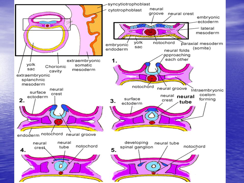
The neural plate folds in upon itself to form the neural tube, which will later differentiate into the spinal cord and the brain, eventually forming the central nervous system.

4
Different portions of the neural tube form by two different processes, called primary and secondary neurulation, in different species. In primary neurulation, the neural plate creases inward until the edges come in contact and fuse. In secondary neurulation, the tube forms by hollowing out of the interior of a solid precursor.

5
Primary neurulation occurs in response to soluble growth factors secreted by the notochord.
Ectodermal cells are induced to form neuroectoderm from a variety of signals. Ectoderm sends and receives signals of BMP4 (bone morphogenic protein) and cells which receive BMP4 signal develop into epidermis.
 and cells which receive BMP4 signal develop into epidermis.")
6
The inhibitory signals chordin, noggin and follistatin are needed to form neural plate. These inhibitory signals are created and emitted by the spemann organiser. Cells which do not receive bone morphogenic protein (BMP4) signaling due to the effects of the inhibitory signals will develop into the anterior neuroectoderm cells of the neural plate. Cells which receive FGF (fibroblast growth factor) in addition to the inhibitory signals form posterior neural plate cells.
 signaling due to the effects of the inhibitory signals will develop into the anterior neuroectoderm cells of the neural plate. Cells which receive FGF (fibroblast growth factor) in addition to the inhibitory signals form posterior neural plate cells.")
7
Shape Change The cells of the neural plate are signaled to become high-columnar and can be identified through microscopy as different from the surrounding epiblastic ectoderm. The cells move laterally and away from the central axis and change into a truncated pyramid shape. This pyramid shape is achieved through tubulin and actin in the apical portion of the cell which constricts as they move.

8
The variation in cell shapes is partially determined by the location of the nucleus within the cell, causing bulging in areas of the cells forcing the height and shape of the cell to change.

9
TAHAP AWAL PERKEMBANGAN OTAK
Proliferasi sel Perubahan bentuk sel Migrasi sel PEMBENTUKAN NEURAL TUBE DLHPs Kalthoff (2001) ; Wolpert, (2002) PEMBENTUKAN NEURAL PLATE MHPs
 ; Wolpert, (2002) PEMBENTUKAN NEURAL PLATE. MHPs.")
10
Folding The process of the flat neural plate folding into the cylindrical neural tube is termed primary neurulation. As a result of the cellular shape changes, the neural plate forms the medial hinge point (MHP). The expanding epidermis puts pressure on the MHP and causes the neural plate to fold resulting in neural folds and the creation of the neural groove.
. The expanding epidermis puts pressure on the MHP and causes the neural plate to fold resulting in neural folds and the creation of the neural groove.")
13
The neural folds form dorsolateral hinge points (DLHP) and pressure on this hinge causes the neural folds to meet and fuse at the midline. The fusion requires the regulation of cell adhesion molecules. The neural plate switches from E-cadherin expression to N-cadherin and N-CAM expression to recognize each other as the same tissue and close the tube. This change in expression stops the binding of the neural tube to the epidermis.

15
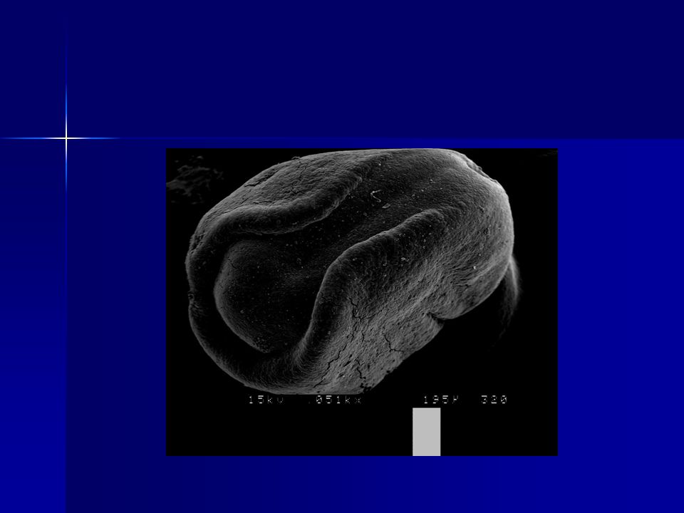
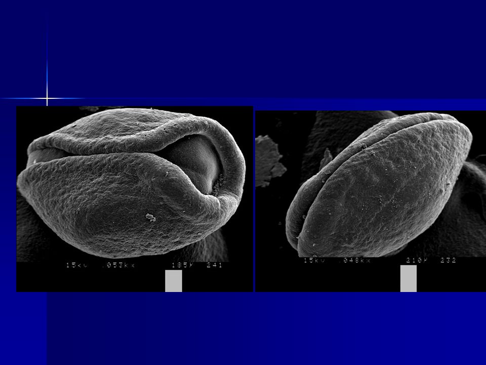
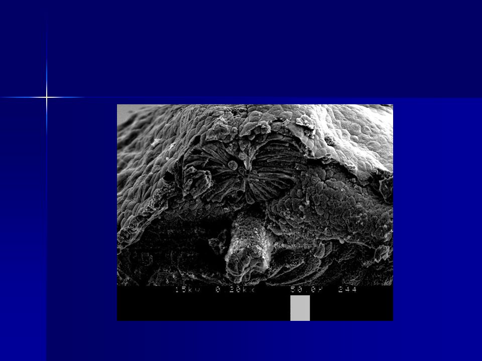
Gambar 5.1. Embrio mencit umur kebuntingan 8,5 hari; a. neural fold; b. neural groove; c. titik fusi pertama pembentukan atap neural tube.

16
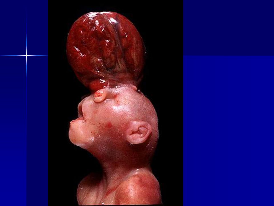
NEURAL TUBE DEFECTS (NTDs)
Eksensefali Spina bifida Normal Eksensefali karena kegagalan penutupan atau reopening neural tube anterior Ada beberapa type eksensefali

17
TYPE EKSENSEFALI (Matsumoto et al., 2002)
")
19
The notochord plays an integral role in the development of the neural tube. Prior to neurulation, during the migration of epiblastic endoderm cells towards the hypoblastic endoderm, the notochordal process opens into an arch termed the notochordal plate and attaches overlying neuroepithelium of the neural plate.

20
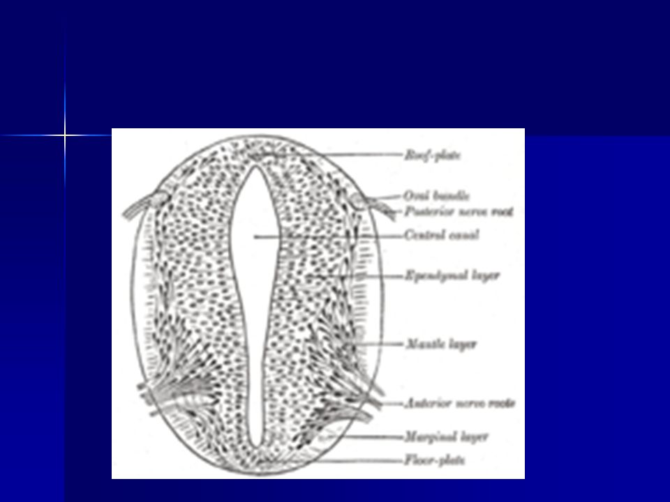
The notochordal plate then serves as an anchor for the neural plate and pushes the two edges of the plate upwards while keeping the middle section anchored. Some of the notochodral cells become incorporated into the center section neural plate to later form the floor plate of the neural tube. The notochord plate separates and forms the solid notochord.

21
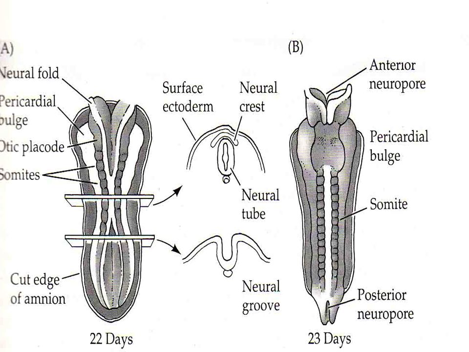
The folding of the neural tube to form an actual tube does not occur all at once.
Instead, it begins approximately at the level of the fourth somite at Carnegie stage 9 (around Embryonic day 20 in humans). The lateral edges of the neural plate touch in the midline and join together. This continues both cranially (toward the head) and caudally (toward the tail). The openings that are formed at the cranial and caudal regions are termed the cranial and caudal neuropores.
. The lateral edges of the neural plate touch in the midline and join together. This continues both cranially (toward the head) and caudally (toward the tail). The openings that are formed at the cranial and caudal regions are termed the cranial and caudal neuropores.")
23
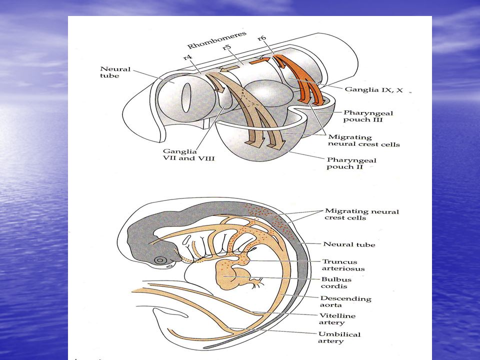
Bakal bumbung neural system saraf pusat
Ectoderm Bakal pial neural sist. saraf perifer , ganglion medulla adrenal, sel – sel pigmen, rawan larink dan kepala Bakal epidermis Penebalan epidermis (lensa mata, telinga dalam, putting – putting pengecap) Kulit, rambut, tanduk , kuku, lapisan permukaan mulut dan anus, hipofisa anterior.
 Kulit, rambut, tanduk , kuku, lapisan permukaan mulut. dan anus, hipofisa anterior.")
24
(Memanjang dan menebal)
Neurulasi Ectoderm Diinduksi Terbentuknya notocord Neural plate (Memanjang dan menebal) tepi neural plate menebal dan tumbuh keatas Neural fold (lipatan neural) dan neural grove (parit neural)
 tepi neural plate menebal. dan tumbuh keatas. Neural fold (lipatan neural) dan neural grove (parit neural)")
25
neural tube neural crest
n. fold tumbuh, saling mendekati daerah dorsal tengah dan akhirnya melebur neural tube neural crest migrasi seluruh tubuh system saraf pusat sel – sel pigmen, sistem saraf tepi dan medula adrenal

28
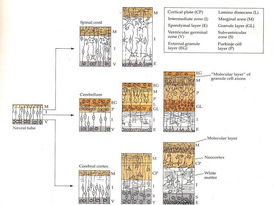
Diferensiasi Bumbung neural berlangsung melalui tiga cara secara serentak, yaitu ;
Secara anatomi ; bumbung neural dan rongganya menggelembung, berkontraksi sehingga terbentuk ruang – ruang (ventrikel otak dan sunsum tulang belakang)
")
29
Tingkat jaringan ; sel –sel dinding. bumbung neural menyusun diri
Tingkat jaringan ; sel –sel dinding bumbung neural menyusun diri sehingga membentuk bagian – bagian fungsional khusus dari otak dan medula spinalis Tingkat selular ; sel –sel akan berdiferentsiasi menjadi berbagai neuron dan sel – sel penunjangnya (sel glial/sel astrosit).
.")
30
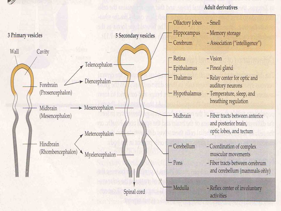
Pada awalnya neural tube berbentuk lurus
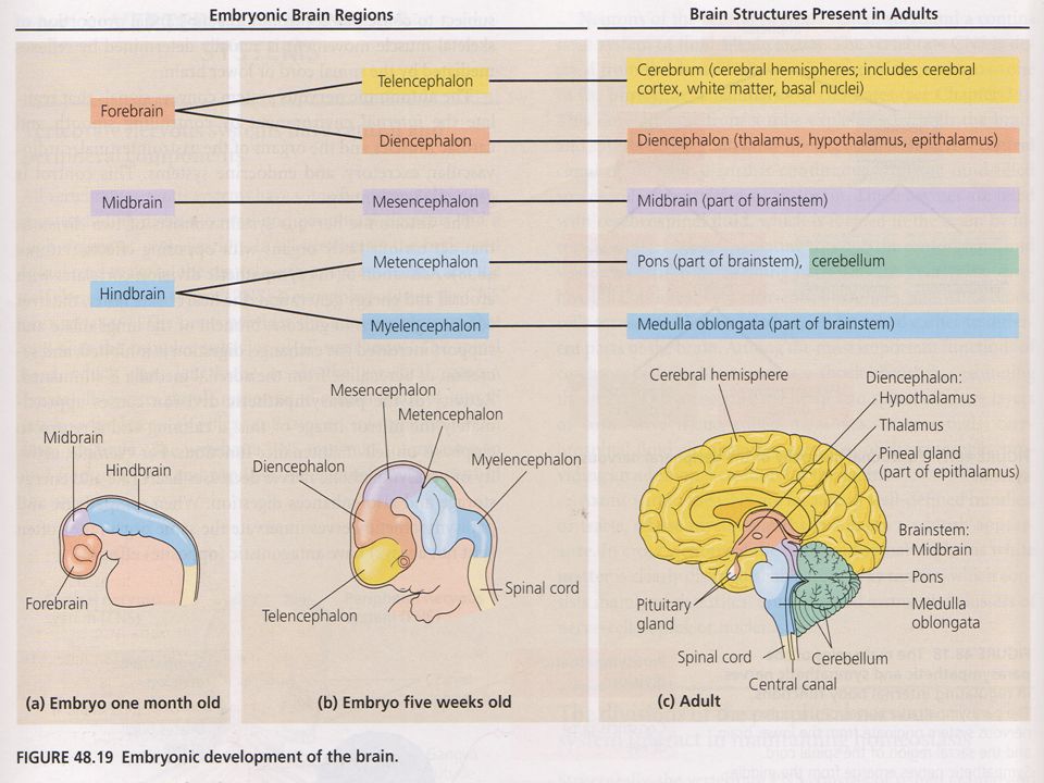
Sebelum neural tube posterior terbentuk, neural tube anterior telah memulai pembentukan otak. Neural tube menggelembung membentuk 3 vesikula yaitu; # Prosencephalon (otak depan) # Mesencephalon (otak tengah) #Rhombencephalon (otak belakang) Pada waktu ujung posterior neural tube menutup, dibentuk penonjolan baru, vesikula optik, menonjol dari kedua sisi lateral otak depan. Pada aves dan mamalia pembatas pada ketiga wilayah otak tampak lebih jelas karena terjadi pembengkokan (fleksi) pada daerah bakal otak yaitu fleksi kranealis dan servikalis.
 # Mesencephalon (otak tengah) #Rhombencephalon (otak belakang) Pada waktu ujung posterior neural tube menutup, dibentuk penonjolan baru, vesikula optik, menonjol dari kedua sisi lateral otak depan. Pada aves dan mamalia pembatas pada ketiga wilayah otak tampak lebih jelas karena terjadi pembengkokan (fleksi) pada daerah bakal otak yaitu fleksi kranealis dan servikalis.")
31
Pada waktu ujung posterior neural tube menutup, dibentuk penonjolan baru, vesikula optik, menonjol dari kedua sisi lateral otak depan, sebagai calon mata. Pada aves dan mamalia calon otak menjadi tiga wilayah otak tampak lebih jelas karena terjadi pembengkokan (fleksi) pada daerah bakal otak yaitu fleksi kranealis dan servikalis.
 pada daerah bakal otak yaitu fleksi kranealis dan servikalis.")
39
Perkembangan otak selanjutnya
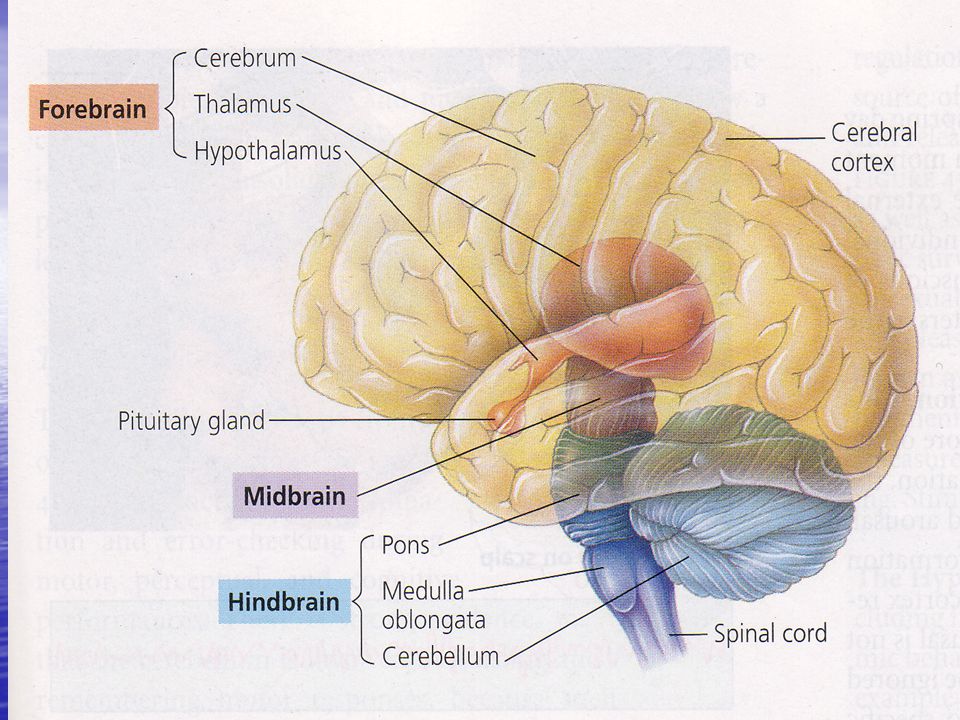
Telencephalon cerebrum (Anterior) (otak besar) 1. Prosencephalon Diencephalon (caudal) daerah thalamus, hipotalamus dengan hipofisa dan hipofisa posterior 2. Mesencephalon Rongga aquaduct cerebral
 (otak besar) 1. Prosencephalon. Diencephalon. (caudal) daerah thalamus, hipotalamus. dengan hipofisa dan hipofisa posterior. 2. Mesencephalon Rongga aquaduct cerebral.")
40
Metencephalon (anterior) Cerebrum, Pons varoli 3. Rhombencephalon Myencephalon (posterior) Medulla oblongata
 Medulla oblongata.")
42
Tahapan dari Siklus Sel

47
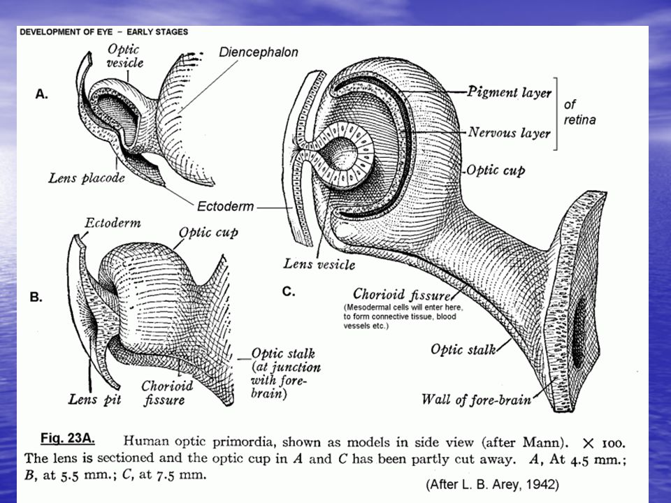
Organogenesis mata Prosencephalon vesicle optic optic cup
Optic cup terdiri 2 lapisan : retina (terdapat sel-sel sensoris) tapetum nigrum (berpigmen) Retina terdiri dari lapisan inti luar, lapisan inti dalam dan lapisan sel ganglion Ektoderm di depan optic cup penebalan placode lensa
 tapetum nigrum (berpigmen) Retina terdiri dari lapisan inti luar, lapisan inti dalam dan lapisan sel ganglion. Ektoderm di depan optic cup penebalan. placode lensa.")
48
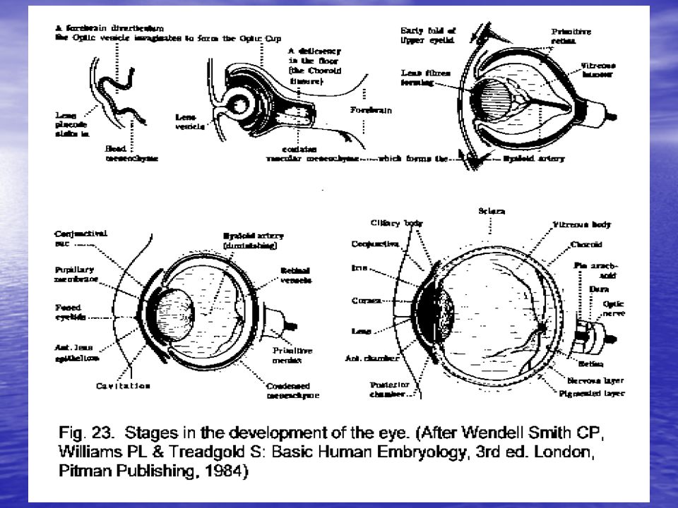
Pembentukan Mata

Presentasi serupa


>")


