Upload presentasi
1
Copyright © 2008 Pearson Education, Inc., publishing as Benjamin Cummings

2
SYSTEMA CIRCULATORIUS KLASIFIKASI: SISTEM KARDIOVASKULAR PEMBULUH DARAH JANTUNG SISTEM LIMFATIK NODUS LYMPHATICUS PEMBULUH LIMFE HUBUNGAN STRUKTURAL/FUNGSIONAL: SISTEM KARDIOVASKULAR BERHUBUNGAN DENGAN SISTEM LIMFATIK HISTOLOGI: SELURUH SISTEM DIBATASI OLEH ENDOTEL

3
Copyright © 2008 Pearson Education, Inc., publishing as Benjamin Cummings HUBUNGAN TIMBAL BALIK SISTEM LIMFATIK DAN SISTEM KARDIOVASKULER SISTEM LIMFATIK SISTEM KARDIO-VASKULER DUCTUS THORACICUS ET DUCTUS LYMPHATICUS DEXTER BERMUARA DALAM VENA BESAR SEBAGAI BAGIAN SISTEM PEMBULUH DARAH SISTEM KARDIOVASKULER SISTEM LIMFATIK CAIRAN LIMFE DENGAN LIMFOSIT DALAM VENULA POST-CAPILER DALAM SETIAP NODUS LYMPHATICUS SEBAGAI BAGIAN DARI SISTEM PEMBULUH DARAH, MASUK SISTEM LIMFATIK, YANG PADA AKHIRNYA DITAMPUNG DALAM KEDUA SALURAN LIMFE BESAR

4
Copyright © 2008 Pearson Education, Inc., publishing as Benjamin Cummings SYSTEMA CIRCULATORIUS JANTUNG ARTERIA BESAR ARTERIA SEDANG ARTERIA KECIL PEMBULUH KAPILER VENA KECIL VENA SEDANG VENA BESAR SISTEM LIMFATIKA DUCTUS THORACICUS SISTEMSISTEM KARDIOVASKULAR KARDIOVASKULARSISTEMSISTEM KARDIOVASKULAR KARDIOVASKULAR

5
Copyright © 2008 Pearson Education, Inc., publishing as Benjamin Cummings SISTEM LIMFATIK KAPILER LIMFE BUNTU (vasa aferentia) (vasa aferentia) NODUS LYMPHATICUS PEMBULUH LIMFE BESAR PEMBULUH DARAH VENA JANTUNG Vasa eferentia DUCTUS THORACICUS ET DUCTUS LYMPHATICUS DEXTER
 (vasa aferentia) NODUS LYMPHATICUS PEMBULUH LIMFE BESAR PEMBULUH DARAH VENA JANTUNG Vasa eferentia DUCTUS THORACICUS ET DUCTUS LYMPHATICUS DEXTER")
6
Copyright © 2008 Pearson Education, Inc., publishing as Benjamin Cummings ALIRAN CAIRAN LIMFE CAIRAN LIMFE (CAIRAN JARINGAN) PLASMA LIMFOSIT PEMBULUH LIMFE DIMULAI DENGAN KAPILER LIMFE BUNTU MENAMPUNG DARI CAIRAN JARINGAN NODUS LYMPHATICUS MENAMPUNG KAPILER PADA PERMUKAAN CEMBUNG PEMBULUH LIMFE LEBIH BESAR MENAMPUNG DARI VASA EFERENTIA N. LYMPHATICUS PEMBULUH LIMFE BESAR MENUJU KE JANTUNG DIAMETER PEMBULUH LIMFE SEMAKIN BESAR DUCTUS THORACICUS V. SUBCLAVIA SINISTRA DUCTUS LYMPHATICUS DEXTER V. SUBCLAVIA DEXTRA

7
Copyright © 2008 Pearson Education, Inc., publishing as Benjamin Cummings ALIRAN CAIRAN LIMFE NODUS LYMPHATICUS ARTERI VENA

8
Copyright © 2008 Pearson Education, Inc., publishing as Benjamin Cummings SIRKULASI PLASMA DAN LIMFOSIT SISTEM LIMFE SISTEM KARDIO - VASKULER

9
Copyright © 2008 Pearson Education, Inc., publishing as Benjamin Cummings DINDING VASA LYMPHATICA VASA AFERENTIA MENAMPUNG CAIRAN JARINGAN DIAMETER LEBIH BESAR SEDIKIT DARI KAPILER DARAH SELAPIS SEL ENDOTEL TIPIS TIDAK ADA PERISIT MASUK KE DALAM SINUS LYMPHATICUS DALAN NODUS LYMPHATICUS KELUAR DARI HILUS SEBAGAI VASA EFERENTIA STRUKTUR DINDING SAMA DENGAN VASA AFERENTIA MENGANGKUT CAIRAN LIMFE DENGAN LIMFOSIT VASA LYMPHATICA MENERIMA BEBERAPA VASA EFERENTIA, DIAMETER BERTAMBAH BESAR DINDING BERTAMBAH TEBAL DI BAGIAN DALAM DILENGKAPI DENGAN VALVULA DUCTUS THORACICUS DAN DUCTUS LYMPHATICUS DEXTER

10
Copyright © 2008 Pearson Education, Inc., publishing as Benjamin Cummings DINDING VASA LYMPHATICA VENULA VALVULA

11
Copyright © 2008 Pearson Education, Inc., publishing as Benjamin Cummings VASA LYMPHATICA BESAR VASA LYMPHATICA BERDIAMETER >0,2 mm DILENGKAPI VALVULA DIBEDAKAN 3 LAPISAN DINDING: TUNICA INTIMA TUNICA MEDIA 2 LAPISAN SEL-SEL OTOT POLOS TUNICA ADVENTITIA BANYAK MENGANDUNG SERABUT KOLAGEN DAN ELASTIS DUCTUS THORACICUS ET DUCTUS LYMPHATICUS DEXTER (PEMBULUH LIMFE TERBESAR) DILENGKAPI DENGAN VALVULA 3 LAPISAN DINDING YANG KURANG JELAS TUNICA INTIMA: ENDOTIL DAN SERABUT KOLAGEN & ELAS TUNICA MEDIA : SEL OTOT POLOS TUNICA ADVENTITIA: SEL-SEL OTOT POLOS MEMANJANG
 DILENGKAPI DENGAN VALVULA 3 LAPISAN DINDING YANG KURANG JELAS TUNICA INTIMA: ENDOTIL DAN SERABUT KOLAGEN & ELAS TUNICA MEDIA : SEL OTOT POLOS TUNICA ADVENTITIA: SEL-SEL OTOT POLOS MEMANJANG")
12
Copyright © 2008 Pearson Education, Inc., publishing as Benjamin Cummings VASA LYMPHATICA BESAR DUCTUS THORACICUS DUCTUS LYMPHATICUS DEXTER VENA CAVA SUPERIOR

13
Copyright © 2008 Pearson Education, Inc., publishing as Benjamin Cummings Arteries Elastic arteries Muscular arteries Arterioles Capillaries Continuous Fenestrated Sinusoidal Veins Veins, venules

14
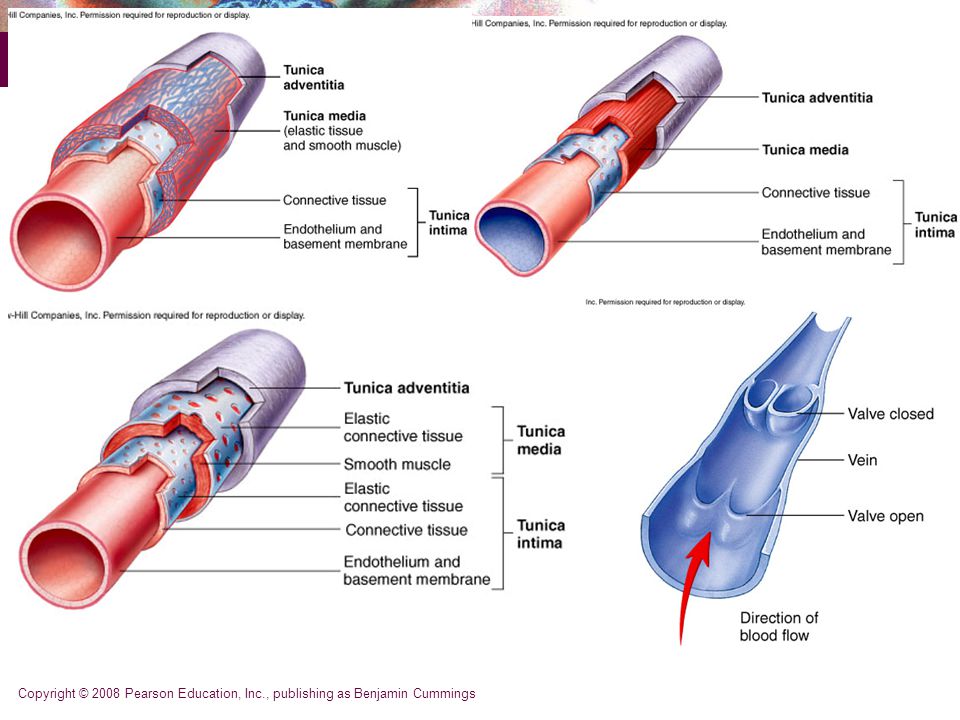
Copyright © 2008 Pearson Education, Inc., publishing as Benjamin Cummings Vessel Structure - General All vessels same basic structure 3 wall layers (or tunics) Tunica adventitia (externa) - elastic and laminar fibers Tunica media thickest layer elastic fibers and smooth muscle fibers Tunica interna (intima) endothelium – non-stick layer basement membrane internal elastic lamina Lumen - opening
 Tunica adventitia (externa) - elastic and laminar fibers Tunica media thickest layer elastic fibers and smooth muscle fibers Tunica interna (intima) endothelium – non-stick layer basement membrane internal elastic lamina Lumen - opening")
15
Copyright © 2008 Pearson Education, Inc., publishing as Benjamin Cummings Structure of Blood Vessels Composed of three layers (tunics) Tunica intima – composed of simple squamous epithelium Tunica media – sheets of smooth muscle Contraction – vasoconstriction Relaxation – vasodilation Tunica externa – composed of connective tissue Lumen Central blood-filled space of a vessel
 Tunica intima – composed of simple squamous epithelium Tunica media – sheets of smooth muscle Contraction – vasoconstriction Relaxation – vasodilation Tunica externa – composed of connective tissue Lumen Central blood-filled space of a vessel")
16
Copyright © 2008 Pearson Education, Inc., publishing as Benjamin Cummings

18
Structure of Arteries, Veins, and Capillaries Figure 19.1a

19
Copyright © 2008 Pearson Education, Inc., publishing as Benjamin Cummings Types of Blood Vessels Arteries – carry blood away from the heart Capillaries – smallest blood vessels The site of exchange of molecules between blood and tissue fluid Veins – carry blood toward the heart

20
Copyright © 2008 Pearson Education, Inc., publishing as Benjamin Cummings

21
Types of Arteries Elastic arteries – the largest arteries Diameters range from 2.5 cm to 1 cm Includes the aorta and its major branches Sometimes called conducting arteries High elastin content dampens surge of blood pressure Aorta, brachiocephalic, common carotid, subclavian, vertebral, pulmonary, common iliac

22
Copyright © 2008 Pearson Education, Inc., publishing as Benjamin Cummings Elastic (conducting) arteries Near heart Thick walls More elastic fiber, less smooth muscle Lose elasticity with aging Vessel Structure – Elastic Arteries
 arteries Near heart Thick walls More elastic fiber, less smooth muscle Lose elasticity with aging Vessel Structure – Elastic Arteries")
23
Copyright © 2008 Pearson Education, Inc., publishing as Benjamin Cummings Vessel Structure - Elastic Arteries Aorta and elastic arteries Can vasoconstrict or vasodilate Large arteries expand, absorb pressure wave then release it with elastic recoil - Windkessel effect Help to push blood along during diastole With aging have less expansion and recoil

24
Copyright © 2008 Pearson Education, Inc., publishing as Benjamin Cummings Types of Arteries Muscular (distributing) arteries Lie distal to elastic arteries Capable of grater vasoconstriction and vaodilation to adjust the blood flow Diameters range from 1 cm to 0.3 mm Includes most named arteries Tunica media is thick, More smooth muscle Less elastic fibers Many of the arteries anastomose Unique features Internal and external elastic laminae Distribute blood to skeletal muscles & internal organs Ex: external carotid, brachial, mesenteric, femoral Figure 19.2b
 arteries Lie distal to elastic arteries Capable of grater vasoconstriction and vaodilation to adjust the blood flow Diameters range from 1 cm to 0.3 mm Includes most named arteries Tunica media is thick, More smooth muscle Less elastic fibers Many of the arteries anastomose Unique features Internal and external elastic laminae Distribute blood to skeletal muscles & internal organs Ex: external carotid, brachial, mesenteric, femoral Figure 19.2b")
25
Copyright © 2008 Pearson Education, Inc., publishing as Benjamin Cummings Figure 19.2c Types of Arteries Arterioles Smallest arteries Diameters range from 0.3 mm to 10 µm Larger arterioles possess all three tunics Diameter of arterioles controlled by Local factors in the tissues (0 2 levels) Sympathetic nervous system hormonal stimulation
 Sympathetic nervous system hormonal stimulation")
26
Copyright © 2008 Pearson Education, Inc., publishing as Benjamin Cummings Arterioles Arterioles are small arteries that deliver blood to capillaries. Also have the three layers as an artery. Tunica media 1-2 layers of smooth muscle fibers A change in diameter of arterioles can significantly affect blood pressure. Through constriction and dilation, arterioles assume a key role in regulating blood flow from arteries into capillaries and in altering arterial blood pressure.

27
Copyright © 2008 Pearson Education, Inc., publishing as Benjamin Cummings Vessel Structure - Capillaries Allow exchange of nutrients and wastes between blood and tissue Capillary structure - simple Basal lamina - connective tissue Endothelial cells Structure/function

28
Copyright © 2008 Pearson Education, Inc., publishing as Benjamin Cummings Arteriole: structure 1. Metarteriole 2. Arteriole 3. Capillary Activity 3

29
Copyright © 2008 Pearson Education, Inc., publishing as Benjamin Cummings Capillaries Smallest blood vessels Diameter from 8–10 µm Red blood cells pass through single file Site-specific functions of capillaries Lungs – oxygen enters blood, carbon dioxide leaves Small intestines – receive digested nutrients Endocrine glands – pick up hormones Kidneys – removal of nitrogenous wastes

30
Copyright © 2008 Pearson Education, Inc., publishing as Benjamin Cummings Capillaries Tempat pertukaran material nutrien antara darah dan jaringan. Tempat terjadinya mikrosirkulasi:aliran darah dari arteriole menuju venule malaui kapiler. capiller tidak berpori (continuous Capillaries) dan berpori ( fenestrated). Precappilary sphincters mengatur aliran darah melalui capillaries. Pada liver berupa sinusoid
 dan berpori ( fenestrated). Precappilary sphincters mengatur aliran darah melalui capillaries. Pada liver berupa sinusoid.")
31
Copyright © 2008 Pearson Education, Inc., publishing as Benjamin Cummings Capillaries Capillary walls are made of a single layer of endothelial cells and a basement membrane. They have no tunica media or tunica externa. Body tissues with high metabolic requirements, such as muscles, kidneys, liver and nervous system, have an extensive network of capillaries. Tissues with low metabolic requirements have fewer capillaries-tendons and ligaments. All covering and lining epithelia, cornea and lens of the eye-lack capillaries.

32
Copyright © 2008 Pearson Education, Inc., publishing as Benjamin Cummings Types of Capillaries True capillaries:emerge from arterioles and metarterioles. Continuous capillaries-found in skeletal and smooth muscle, connective tissues and the lungs. Fenestrated capillaries-kidneys, villi os the SI, choroid plexuses in brain, ciliary process, endocrine glands. Sinusoids:are wider and more winding than other capillaries. Present in liver, red bone marrow, pleen, ant.pit. Gland, and parathyroid glands.

33
Copyright © 2008 Pearson Education, Inc., publishing as Benjamin Cummings RBCs in a Capillary Figure 19.3

34
Copyright © 2008 Pearson Education, Inc., publishing as Benjamin Cummings Capillary Beds Network of capillaries running through tissues Precapillary sphincters Regulate the flow of blood to tissues Tendons and ligaments – poorly vascularized Epithelia and cartilage – avascular Receive nutrients from nearby CT

35
Copyright © 2008 Pearson Education, Inc., publishing as Benjamin Cummings Capillary Beds Figure 19.4a

36
Copyright © 2008 Pearson Education, Inc., publishing as Benjamin Cummings Capillary Beds Figure 19.4b

37
Copyright © 2008 Pearson Education, Inc., publishing as Benjamin Cummings

38
Local control of blood vessels Sphincters contract or relax based on demand for: nutrients (AA, glucose, fatty acids) Dissolved gases (O 2, CO 2 load, lactic acid) Additional capillaries grow in to area to satisfy increased energy demands
 Dissolved gases (O 2, CO 2 load, lactic acid) Additional capillaries grow in to area to satisfy increased energy demands")
39
Copyright © 2008 Pearson Education, Inc., publishing as Benjamin Cummings Capillary Permeabillity Endothelial cells – held together by tight junctions and desmosomes Intercellular clefts – gaps of unjoined membrane Small molecules can enter and exit Two types of capillary Continuous – most common Fenestrated – have pores Sinusoid

40
Copyright © 2008 Pearson Education, Inc., publishing as Benjamin Cummings Types of Capillaries 3 types of capillaries 1. Continuous capillaries continuous endothelial cells except for cleft between cells tight junctions between endothelial cells prevent most things from leaving caps most capillaries in body

41
Copyright © 2008 Pearson Education, Inc., publishing as Benjamin Cummings Structure of Capillaries – Cross Section Figure 19.5a

42
Copyright © 2008 Pearson Education, Inc., publishing as Benjamin Cummings Types of Capillaries 2. Fenestrated capillaries fenestrations (slits) allow for filtration of small substances glomerular capillaries in kidney
 allow for filtration of small substances glomerular capillaries in kidney.")
43
Copyright © 2008 Pearson Education, Inc., publishing as Benjamin Cummings Structure of Capillaries – Cross Section Figure 19.5b

44
Copyright © 2008 Pearson Education, Inc., publishing as Benjamin Cummings Routes of Capillary Permeability Four routes into and out of capillaries Direct diffusion Through intercellular clefts Through cytoplasmic vesicles Through fenestrations

45
Copyright © 2008 Pearson Education, Inc., publishing as Benjamin Cummings Low Permeability Capillaries Blood-brain barrier Capillaries have complete tight junctions No intercellular clefts are present Vital molecules pass through Highly selective transport mechanisms Not a barrier against Oxygen, carbon dioxide, and some anesthetics

46
Copyright © 2008 Pearson Education, Inc., publishing as Benjamin Cummings Types of Capillaries 3. Sinusoid capillaries wider gaps between endothelial cells allowing RBC’s to exit the caps found in liver

47
Copyright © 2008 Pearson Education, Inc., publishing as Benjamin Cummings Sinusoids Wide, leaky capillaries found in some organs Usually fenestrated Intercellular clefts are wide open Occur in bone marrow and spleen Sinusoids have a large diameter and twisted course

48
Copyright © 2008 Pearson Education, Inc., publishing as Benjamin Cummings Sinusoids Figure 19.5c

49
Copyright © 2008 Pearson Education, Inc., publishing as Benjamin Cummings Veins Conduct blood from capillaries toward the heart Blood pressure is much lower than in arteries Smallest veins – called venules Diameters from 8 – 100 µm Smallest venules – called postcapillary venules Venules join to form veins Tunica externa is the thickest tunic in veins

50
Copyright © 2008 Pearson Education, Inc., publishing as Benjamin Cummings Vessel Structure – Veins Veins Interna thicker than arteries Media thinner, less muscle Externa thick Valves Pressure low High compliance - change volume easily with small change in pressure Varicose veins

51
Copyright © 2008 Pearson Education, Inc., publishing as Benjamin Cummings CIRI MENGANGKUT DARAH KE JANTUNG JUMLAH LEBIH BESAR DARIPADA ARTERIA MENDEKATI JANTUNG DIAMETER MAKIN BESAR BIASANYA BERADA DI DEKAT ARTERINYA KETEBALAN DINDING LEBIH TIPIS DENGAN VALVULA BIASANYA PADA SEDIAAN DALAM KONDISI KOLAPS KLASIFIKASI: VENA BESAR VENA SEDANG VENA KECIL = VENULA DINDING TUNICA INTIMA TUNICA MEDIA TUNICA ADVENTITIA VENA

52
Copyright © 2008 Pearson Education, Inc., publishing as Benjamin Cummings VENA BESAR MIKROSKOPIS TUNICA INTIMA ( 45 m - 68 m) ENDOTEL JARINGAN PENGIKAT SANGAT TIPIS TUNICA MEDIA TIDAK BERKEMBANG DENGAN BAIK SERINGKALI TIDAK ADA TUNICA ADVENTITIA MERUPAKAN BAGIAN UTAMA DARI DINDING JARINGAN PENGIKAT: SERABUT ELASTIS DAN SERABUT KOLAGEN YANG MEMANJANG TERUTAMA MENGANDUNG SERABUT OTOT POLOS MEMANJANG CONTOH: VENA CAVA, VENA PORTAE, V. LIENALIS.

53
Copyright © 2008 Pearson Education, Inc., publishing as Benjamin Cummings VENA BESAR OTOT POLOS TUNICA MEDIA CUKUP TEBAL

54
Copyright © 2008 Pearson Education, Inc., publishing as Benjamin Cummings VENA SEDANG (2 - 9 mm) MIKROSKOPIS TUNICA INTIMA (TIPIS) SEL ENDOTEL JARINGAN PENGIKAT TIPIS SEDIKIT SERABUT ELASTIS TUNICA MEDIA (LEBIH TIPIS DARIPADA ARTERI SEDANG) TERUTAMA SEL OTOT POLOS SIRKULER OTOT POLOS DIPISAHKAN SER. KOLAGEN MEMANJANG SEDIKIT FIBROBLAS TUNICA ADVENTITIA (LEBIH TEBAL DARIPADA TUNICA MEDIA) JARINGAN PENGIKAT LONGGAR DENGAN BERKAS TEBAL SERABUT KOLAGEN MEMANJANG DAN ANYAMAN SERABUT ELASTIS BAGIAN DALAM SERING ADA BERKAS SEL-SEL OTOT POLOS MEMANJANG
 JARINGAN PENGIKAT LONGGAR DENGAN BERKAS TEBAL SERABUT KOLAGEN MEMANJANG DAN ANYAMAN SERABUT ELASTIS BAGIAN DALAM SERING ADA BERKAS SEL-SEL OTOT POLOS MEMANJANG.")
55
Copyright © 2008 Pearson Education, Inc., publishing as Benjamin Cummings VENA SEDANG (2 - 9 mm)
")
56
Copyright © 2008 Pearson Education, Inc., publishing as Benjamin Cummings PERBANDINGAN STRUKTUR DINDING ARTERIA SEDANG DAN VENA SEDANG ARTERIA SEDANG VENA SEDANG

57
Copyright © 2008 Pearson Education, Inc., publishing as Benjamin Cummings VENULA (15 m - 200 m) MENERIMA DARAH DARI KAPILER DINDING: TUNICA INTIMA ENDOTEL JARINGAN PENGIKAT, BEBERAPA SEL OTOT POLOS, MAKIN BESAR DIAMETER: SEL-SEL MAKIN RAPAT TUNICA MEDIA 1 ATAU BEBERAPA LAPIS SEL-SEL OTOT POLOS TUNICA ADVENTITIA FIBROBLAS DAN SERABUT TIPIS ELASTIS DAN KOLAGEN MEMANJANG SIFAT: PERMEABILITAS CUKUP TINGGI
 MENERIMA DARAH DARI KAPILER DINDING: TUNICA INTIMA ENDOTEL JARINGAN PENGIKAT, BEBERAPA SEL OTOT POLOS, MAKIN BESAR DIAMETER: SEL-SEL MAKIN RAPAT TUNICA MEDIA 1 ATAU BEBERAPA LAPIS SEL-SEL OTOT POLOS TUNICA ADVENTITIA FIBROBLAS DAN SERABUT TIPIS ELASTIS DAN KOLAGEN MEMANJANG SIFAT: PERMEABILITAS CUKUP TINGGI")
58
Copyright © 2008 Pearson Education, Inc., publishing as Benjamin Cummings VENULA DENGAN KATUP (15 m - 200 m) KATUP
 KATUP")
59
Copyright © 2008 Pearson Education, Inc., publishing as Benjamin Cummings ARTERIOLA DAN VENULA MEMBRANA ELASTICA INTERNA

60
Copyright © 2008 Pearson Education, Inc., publishing as Benjamin Cummings Figure 19.6 Mechanisms to Counteract Low Venous Pressure Valves in some veins Particularly in limbs Skeletal muscle pump Muscles press against thin-walled veins force blood back to heart valves prevent back flow

61
Copyright © 2008 Pearson Education, Inc., publishing as Benjamin Cummings Vascular Anastomoses Vessels interconnect to form vascular anastomoses Organs receive blood from more than one arterial source Neighboring arteries form arterial anastomoses Provide collateral channels Veins anastomose more frequently than arteries

62
Copyright © 2008 Pearson Education, Inc., publishing as Benjamin Cummings Vasa Vasorum Tunica externa of large vessels have Tiny arteries, capillaries, and veins Vasa vasorum vessels of vessels Nourish outer region of large vessels Inner half of large vessels receive nutrients from luminal blood

63
Copyright © 2008 Pearson Education, Inc., publishing as Benjamin Cummings Arteries Sympathetic fibers of the ANS innervate vascular smooth muscle. An inc. in sympathetic stimulation typically stimulates the smooth muscle to contract-vasoconstriction. When sympathetic stimulation decreases, or in presence of certain chemicals-NO, K +, H + and lactic acid, they relax-vasodilation.

64
Copyright © 2008 Pearson Education, Inc., publishing as Benjamin Cummings Vessel Structure - Function Capillary Function Site of exchange between blood and tissues Delivery of nutrients and removal of wastes Slow flow allows time for exchange Mechanisms of nutrient exchange Diffusion - O 2, CO 2, glucose, AA's, hormones diffuse down [ ] gradients If lipid soluble, can travel through cell If water soluble, between cells
![Copyright © 2008 Pearson Education, Inc., publishing as Benjamin Cummings Vessel Structure - Function Capillary Function Site of exchange between blood and tissues Delivery of nutrients and removal of wastes Slow flow allows time for exchange Mechanisms of nutrient exchange Diffusion - O 2, CO 2, glucose, AA s, hormones diffuse down [ ] gradients If lipid soluble, can travel through cell If water soluble, between cells](http://images.slideplayer.info/12/3674103/slides/slide_64.jpg "Copyright © 2008 Pearson Education, Inc., publishing as Benjamin Cummings Vessel Structure - Function Capillary Function Site of exchange between blood and tissues Delivery of nutrients and removal of wastes Slow flow allows time for exchange Mechanisms of nutrient exchange Diffusion - O 2, CO 2, glucose, AA s, hormones diffuse down [ ] gradients If lipid soluble, can travel through cell If water soluble, between cells")
65
Copyright © 2008 Pearson Education, Inc., publishing as Benjamin Cummings Capillary Fluid Exchange Forces driving the movement of fluid Hydrostatic pressure capillary (HP c ) Hydrostatic pressure interstitial fluid (HP if ) Osmotic pressure capillary(OP c ) Osmotic pressure interstitial fluid (OP if ) Net filtration pressure (NFP) is a sum of all Fluid movement Fluid filtered and reabsorbed across capillary wall Starling’s law of the capillaries
 Hydrostatic pressure interstitial fluid (HP if ) Osmotic pressure capillary(OP c ) Osmotic pressure interstitial fluid (OP if ) Net filtration pressure (NFP) is a sum of all Fluid movement Fluid filtered and reabsorbed across capillary wall Starling’s law of the capillaries")
66
Copyright © 2008 Pearson Education, Inc., publishing as Benjamin Cummings Capillary Fluid Exchange On average 85% of fluid filtered at arteriole end is reabsorbed at venular end

67
Copyright © 2008 Pearson Education, Inc., publishing as Benjamin Cummings Normal Coronary Artery Cross Section

68
Copyright © 2008 Pearson Education, Inc., publishing as Benjamin Cummings 60% Narrowing of Coronary Artery

69
Copyright © 2008 Pearson Education, Inc., publishing as Benjamin Cummings 90% Blockage of Coronary Artery calcified arearemaining lumen

70
Copyright © 2008 Pearson Education, Inc., publishing as Benjamin Cummings Atherosclerotic Plaque Histology cholesterol crystal (cleft) foam cells
 foam cells")
71
Copyright © 2008 Pearson Education, Inc., publishing as Benjamin Cummings Thrombus Causing MI “Needle-Like” white spots are cholesterol crystals Thrombus ocluding arteryLikely site of plaque rupture

72
Copyright © 2008 Pearson Education, Inc., publishing as Benjamin Cummings Myocardial Infarction Histology necrosed muscle cellsred blood cells

73
Copyright © 2008 Pearson Education, Inc., publishing as Benjamin Cummings Myocardial Infarction Histology normal muscle cells remaining macrophages and the beginnings of scar tissue


 Reka Indera Malis (18) Muhammad Nizar Rahman (29)>")


