Upload presentasi
Presentasi sedang didownload. Silahkan tunggu

1
Cataract Mula Tarigan, SKp PSIK FK USU

2
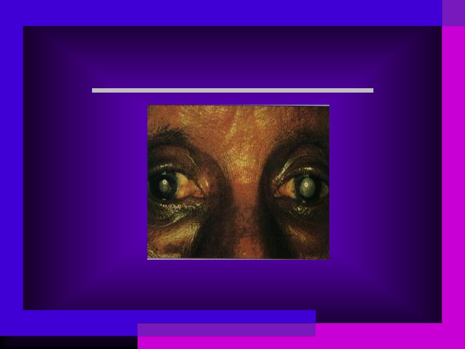
What is a cataract? A cataract is an opacity(or cloudy changes) of the lens that can cause vision problems. Keadaan yang menunjukkan adanya kekeruhan lensa dari yang hanya terbentuk titik sampai kekeruhan lensa yang menyeluruh.
 of the lens that can cause vision problems. Keadaan yang menunjukkan adanya kekeruhan lensa dari yang hanya terbentuk titik sampai kekeruhan lensa yang menyeluruh..")
4
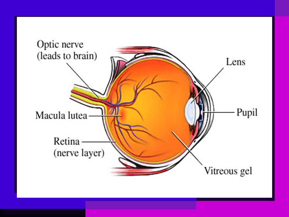
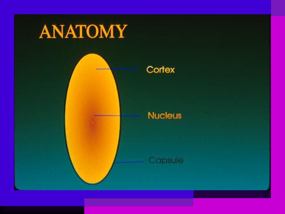
LENS The lens is surrounded by a thick lens capsule which is the basement membrane of the lens epithelial cells. Epithelial cells at the lens equator continue to be produced throughout life,so that older lens fibers are compressed into a central nucleus; younger fibers around the nucleus make up the cortex.

6
causes Aging most common Long-term ultraviolet (UV) light, especially from sunlight Diabetes or other systemic disease Past eye infections, injuries or surgery Smoking Long-term use of certain medications (such as steroids) Heredity
 light, especially from sunlight Diabetes or other systemic disease Past eye infections, injuries or surgery Smoking Long-term use of certain medications (such as steroids) Heredity")
7
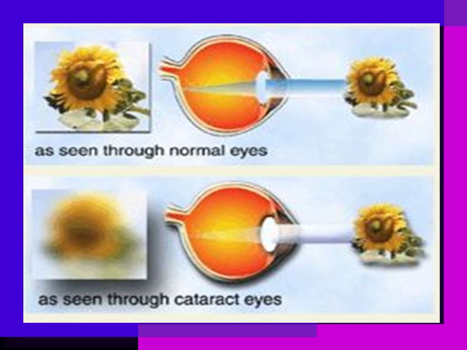
Pathogenesis The lens is made mostly of water and protein. The protein is arranged to let light pass through and focus on the retina. Sometimes some of the protein clumps together. This can start to cloud small areas of the lens, blocking some light from reaching the retina and interfering with vision.

8
normal Vs. cloudy lens

10
Clinical Findings Symptoms Blurring or dimness of vision Colors appear faded Sensitivity to light and glare Double or multiple vision Change in refraction

12
Eye Exam Vision acuity test Slit lamp Ophthalmoscope In most cases, eye drops are used to dilate (widen) pupils before the exam. Tonometry

13
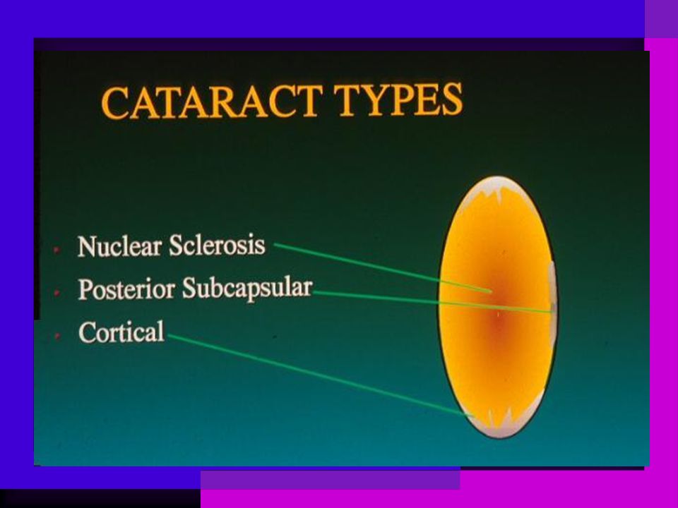
There are three major types of cataract that are named depending on the location within the lens that is most affected. These are cortical, nuclear and posterior subcapsular.

15
Cortical cataract the most common type of age-related cataract four stages as follows:

16
Incipient stage Cortical changes may begin as small peripheral water clefts Radical pattern opacity

17
Intumescent stage The lens takes up water, it becomes intumescent. Anterior chamber gets shallow

18
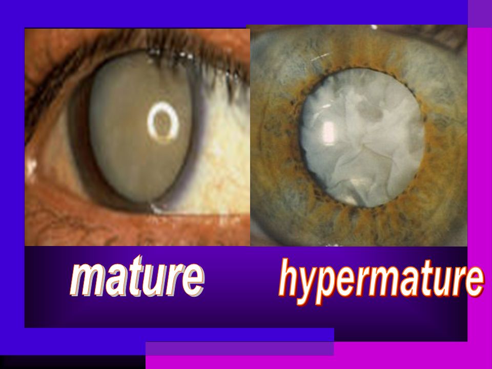
Mature stage Liquid escapes and the lens shrinks The lens protein is totally opaque

19
Hypermature Stage A long-standing or very mature cataract may undergo liquefaction of the lens cortex. This liquid may escape through the intact capsule,leaving a shrunken lens with a wrinkled capsule.

21
This slide shows a lens that has been removed at surgery.

22
Nuclear cataract Early onset (after middle age) The earliest symptom may be improved near vision without glasses ( “ second sight ” ) Other symptoms may include poor hue discrimination or monocular diplopia.
 The earliest symptom may be improved near vision without glasses ( second sight ) Other symptoms may include poor hue discrimination or monocular diplopia.")
23
Posterior subcapsular cataract Located in the cortex near the central posterior capsule It tends to cause visual symptoms earlier in their development owing to involvement of the visual axis. Common symptoms include glare and reduced vision under bright lighting conditions.

24
Congenital Cataract Present at birth or appear shortly thereafter These cataracts may show many different patterns. The opacity may be confined to the area of the embryonic or fetal nucleus with clear cortex surrounding this.

25
Etiology Intra-uterine virus infection Maternal ingestion of Thalidomide, steroids,… Hereditary autosomal dominant recessive X-linked

27
Cataract Treatment Surgery is the only way to remove the cataract. However, if symptoms from a cataract are mild, a change of glasses may be all that is needed for you to function more comfortably. Cataract surgery should be considered when cataracts cause enough loss of vision to interfere with daily activities.

29
ECCE+IOL Extracapsular cataract extraction is a preferred method of cataract surgery It preserves the posterior portion of the lens capsule Posterior chamber IOL can be implanted in the capsular sac

30
Intraocular Lens An IOL is a tiny, transparent, convex lens made of polymer which is inserted in the eye during surgery.

31
IOL 可折式 6mm Foldable IOL

32
Anterior chamber type Posterior chamber type

33
Advantages of IOL Since the lens is placed inside the eye, the patient need not wear glasses for distant vision. Images are clear and of the same dimension without distortion. Full vision is obtained soon after surgery.

34
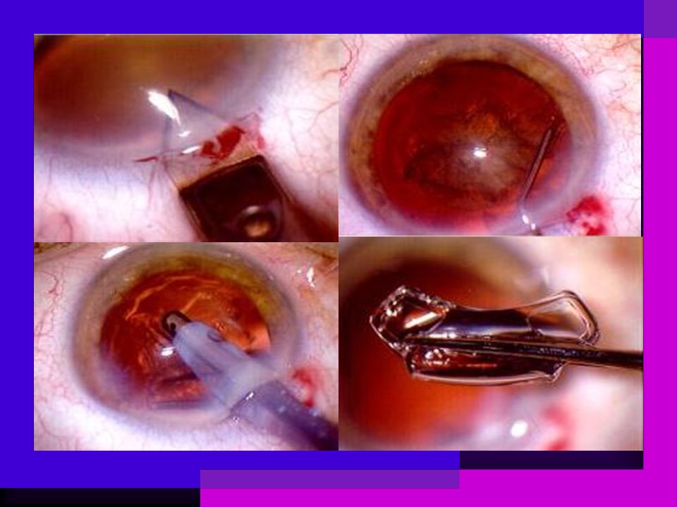
Phacoemulsification Phacoemulsification or phaco refers to ultra-sonic vibration which dissolves the hard nucleus such that the nuclear material and cortex can be aspired through an incision of approximately 3mm. it is the key to advanced, small-incision cataract surgery.

36
Complications posterior capsule opacification cystoid macular edema glaucoma hyphema ptosis infection retinal detachment lens dislocation

39
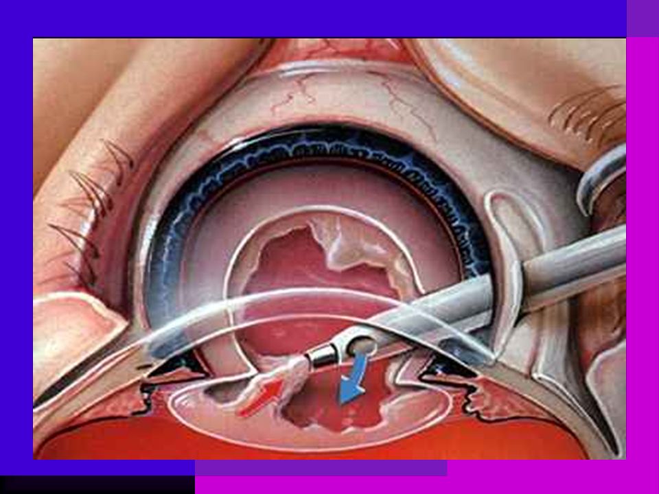
Cataract Surgery animation

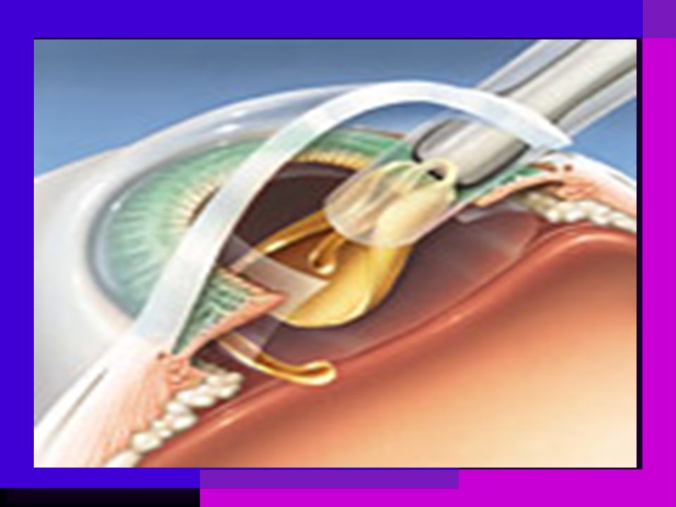
43
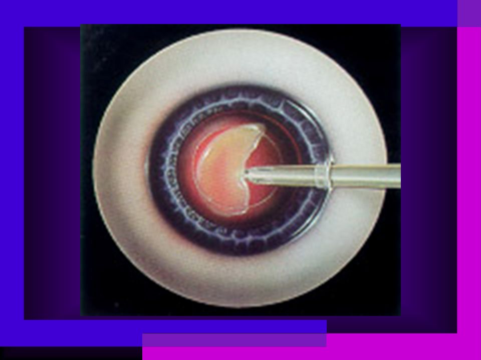
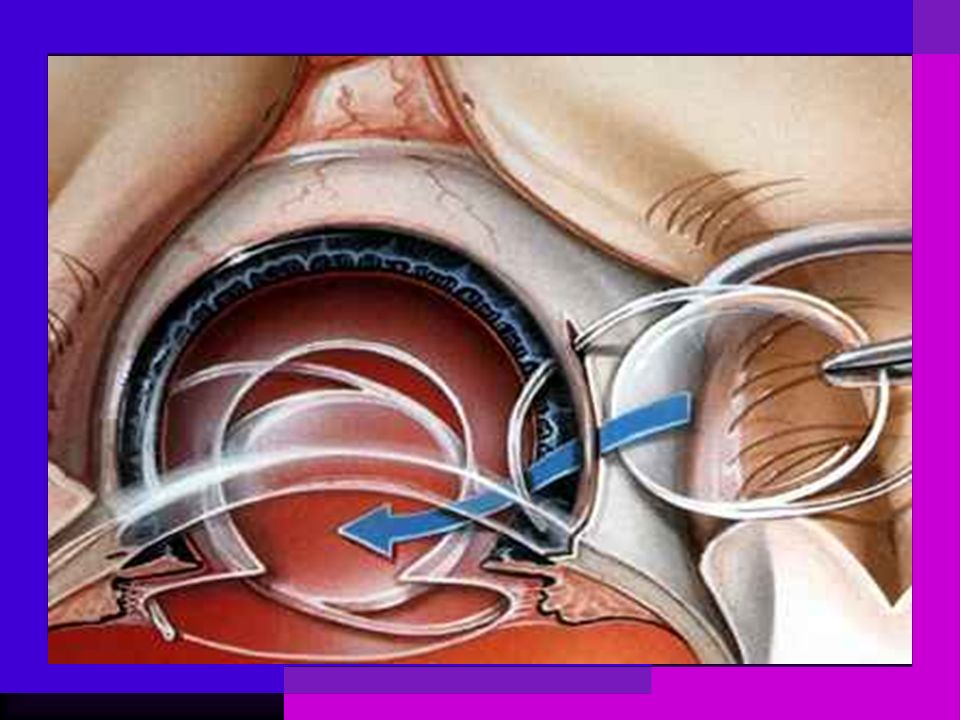
Inserting the new lens

45
Nursing Diagnoses a.Perubahan sensori perseptual: visual b/d kekeruhan pd lensa d/d pupil tampak putih, pasien mengeluhkan pandangan kabur, berkabut, atau pandangan ganda dan gangguan penglihatan. b.Ketakutan/ ansietas b/d kerusakan sensori dan kurang pemahaman mengenai perawatan pasca operasi, pemberian obat. c.Resiko cedera b/d penurunan visus atau berada di lingkungan yang kurang dikenal.

46
d.Resiko cedera b/d komplikasi pasca operasi spt; pendarahan atau peningkatan tekanan intra okuler. e.Defisit perawatan diri b/d kelemahan visual dan perawatan mata pasca operasi. f.Resiko tinggi infeksi b/d prosedur invasif (bedah pengangkatan katarak) g.Kurang pengetahuan ttg kondisi pengobatan dan perawatan pasca operasi b/d terbatasnya informasi atau kesalahan interpretasi informasi.
 g.Kurang pengetahuan ttg kondisi pengobatan dan perawatan pasca operasi b/d terbatasnya informasi atau kesalahan interpretasi informasi..")
47
a.Perubahan sensori perseptual: visual b/d kekeruhan pd lensa d/d pupil tampak putih, pasien mengeluhkan pandangan kabur, berkabut, atau pandangan ganda dan gangguan penglihatan. Tujuan : Pasien mendemonstrasikan peningkatan kemampuan untuk memproses rangsangan visual dan mengkomunikasikan pembatasan pandangan. Kriteria Hasil: –Visus meningkat –Respon verbal peningkatan penglihatan

48
Intervensi Mandiri: 1. kaji ketajaman penglihatan klien 2. berikan pencahayaan yg plg sesuai dgn klien 3. cegah glare atau sinar yg menyilaukan 4. letakkan brg2 pd tempat yang konsisten 5. gunakan materi dgn tulisan besar dan kontras Kolaborasi : pembedahan

49
c. Resiko cedera b/d penurunan visus atau berada di lingkungan yang kurang dikenal. Tujuan: Klien tidak mengalami cedera akibat jatuh. Kriteria Hasil: - Pasien mengenal lingkungan - Pasien tidak jatuh selama perawatan

50
Intervensi: 1.kurangi resiko bahaya dari lingkungan klien. 2.beritahu klien utk mengubah posisi secara perlahan. 3.beritahu klien utk tdk meraih benda untuk stabilitas saat ambulasi. 4.dorong klien utk menggunakan peralatan adaftif (tongkat atau walker) untuk ambulasi sesuai kebutuhan. 5.tekankan pentingnya utk menggunakan pelindung mata saat melakukan aktifitas beresiko tinggi.
 untuk ambulasi sesuai kebutuhan. 5.tekankan pentingnya utk menggunakan pelindung mata saat melakukan aktifitas beresiko tinggi..")
51
g.Kurang pengetahuan ttg kondisi pengobatan dan perawatan pasca operasi b/d terbatasnya informasi atau kesalahan interpretasi informasi. Tujuan : menyatakan pemahaman kondisi/ proses penyakit dan pengobatan. Kriteria Hasil: - Respon verbal memahami proses penyakit dan pengobatan - Menunjukkan tindakan yang kooperatif

52
Intervensi : 1.kaji informasi ttg kondisi individu, prognosis, tipe prosedur atau lensa. 2.tekankan pentingnya evaluasi perawatan rutin. 3.informasikan pasien utk menghindari tetes mata yg dijual bebas. 4.diskusikan kemungkinan efek/interaksi antara obat, mata dan masalah medis pasien. 5.dorong pemasukan cairan adekuat, makanan berserat/kasar, gunakan pelunak feses yg dijual bebas, bila diindikasikan. 6.identifikasi tanda/gejala yg memerlukan upaya evaluasi medis.

53
Warning Signs Reduction in visual acuity Photophobia Purulent discharge ‘ Red Eye ’ Pain vs. ‘ Picking ’

54
Post Operative Requirements Discharge with eyedrops –Dexamethasone –Topical steroid – reduces post-op inflammation Do not lift weights of over 10kg for al least 6 weeks Do not bend from the waist for prolonged periods Wear an eye shield at night for the first 14 days to prevent inadvertent rubbing of the eye when asleep

55
1st day post-op follow up for specific patient groups only: – Glaucoma – Diabetes –Non-standard or complex surgery Review in clinic two weeks after surgery Autorefraction Was the post-op outcome as expected Visual acuity; pathology or refraction?

56
That’s all for today!

Presentasi serupa


>")

 : ADALAH CABANG MATEMATIKA YANG MEMPELAJARI PENGATURAN OBJEK- OBJEK. ADALAH CABANG.>")
