Upload presentasi
Presentasi sedang didownload. Silahkan tunggu

1
Kontrol Terhadap Fungsi Pencernaan
dr. Nuraiza Meutia,M.Biomed Departemen Fisiologi FK USU

2
Aktivitas saluran cerna untuk menjalankan proses percernaan diatur oleh sistem saraf dan endokrin.
Saluran cerna memiliki kemandirian untuk kedua sistem tersebut. Terjadi aktivitas terintegrasi antara kedua sistem mengatur aktivitas motorik dan sekretorik

3
Pada akhir perkuliahan, anda harus dapat :
Menjelaskan anatomi fungsional dinding GIT Menjelaskan aktivitas utama GIT Menjelaskan persarafan otonom yang mengatur GIT Menjelaskan letak dan fungsi pleksus saraf di GIT Menyebutkan nama dan efek neurotransmitter yang bekerja di ENS Menjelaskan peran hormon dalam kontrol GIT (nama hormon, sumber, target dan efek) Menjelaskan proses integrasi sistem saraf dan endokrin di GIT Menjelaskan perbedaan refleks lokal dan refleks sentral dalam regulasi kerja GIT Menjelaskan aktivitas listrik pada otot polos GIT Menjelaskan mekanisme kontraksi dan jenis motilitas GIT Menjelaskan contoh-contoh refleks GI Menjelaskan regulasi aktivitas lambung, usus halus, usus besar, pankreas dan kandung empedu.
 Menjelaskan proses integrasi sistem saraf dan endokrin di GIT. Menjelaskan perbedaan refleks lokal dan refleks sentral dalam regulasi kerja GIT. Menjelaskan aktivitas listrik pada otot polos GIT. Menjelaskan mekanisme kontraksi dan jenis motilitas GIT. Menjelaskan contoh-contoh refleks GI. Menjelaskan regulasi aktivitas lambung, usus halus, usus besar, pankreas dan kandung empedu.")
4
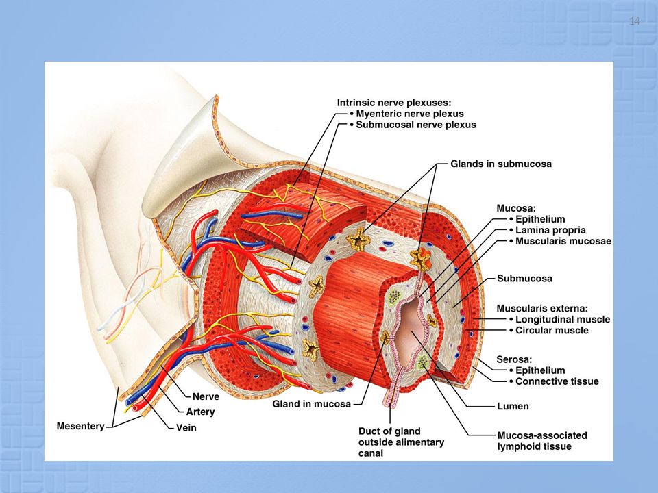
Struktur Dinding GIT

5
Aktivitas GIT : Motilitas Sekresi Digesti Absorbsi Digestive Process

6
Fungsi sistem regulasi di GIT :
Mengatur aktivitas motilitas dan sekresi Mengatur aliran darah ke GIT Menerima dan menyampaikan informasi melalui neuron sensori (aferen) , dari reseptor-reseptor yang menerima stimulus mekanikal, thermal, osmotik dan kimiawi.
 , dari reseptor-reseptor yang menerima stimulus mekanikal, thermal, osmotik dan kimiawi.")
7
GIT NEURAL CONTROL

8
Aktivitas GIT diatur oleh sistem Saraf Otonom
Terdiri dari : - Divisi parasimpatetik - Divisi simpatetik - Enteric Nervous System (ENS)
")
9
Parasimpatetik N. Vagus & N.pelvik
Neuron preganglionik panjang; postganglionik pendek, bersinaps dengan neuron ENS Stimulasi eksitasi aktivitas ENS Mengandung serat sensori aferen (80 %) N.Vagus bersinaps ke neuron ENS di esophagus, lambung,usus halus, sebagian kolon, kandung empedu, & pankreas N.Pelvik bersinaps dengan ENS di usus besar Neurotransmitter : Ach
 N.Vagus bersinaps ke neuron ENS di esophagus, lambung,usus halus, sebagian kolon, kandung empedu, & pankreas. N.Pelvik bersinaps dengan ENS di usus besar. Neurotransmitter : Ach.")
10
Parasympathetic Nervous System Craniosacral

11
Simpatetik Serat simpatetik ke GIT berasal dari medula spinalis segmen T-5 sampai L-2. Neurotransmitter : norepinefrin Aktivitas simpatetik inhibisi motilitas dan sekresi GIT, konstriksi sfinkter dan pembuluh darah.

12
Sympathetic Nervous System Thoracolumbar

13
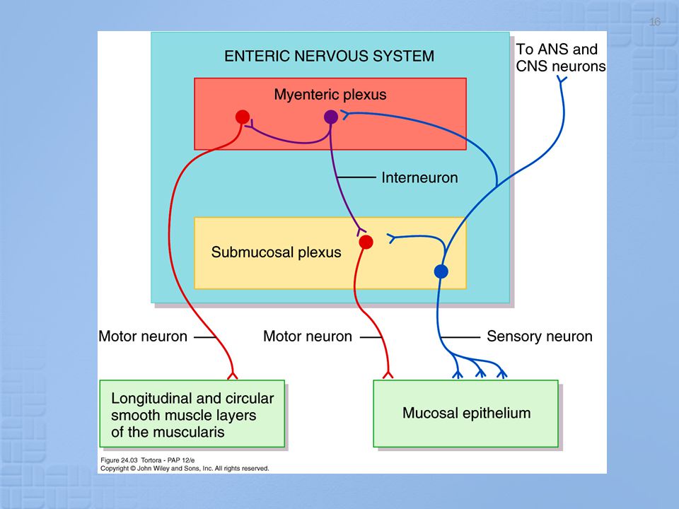
Enteric Nervous System
Enteric Nervous System (ENS) terdapat di seluruh dinding GIT, mulai esophagus sampai anus. Terbentuk dari 100 juta neuron (mengimbangi spinal cord). Memiliki 3 jenis neuron : sensori, motorik, & interneuron ENS tersusun atas 2 pleksus utama : (1) Myenteric plexus atau Auerbach's Plexus: berada di antara lapisan otot sirkular dan longitudinal (outer plexus). Fungsi : mengontrol motilitas GIT (2) Submucosal Plexus atau Meissner's plexus : berada di lapisan submukosa (inner plexus). Fungsi : mengatur sekresi dan aliran darah lokal, sensing perubahan lumen, dan gerak pelipatan mukosa.
 terdapat di seluruh dinding GIT, mulai esophagus sampai anus. Terbentuk dari 100 juta neuron (mengimbangi spinal cord). Memiliki 3 jenis neuron : sensori, motorik, & interneuron. ENS tersusun atas 2 pleksus utama : (1) Myenteric plexus atau Auerbach s Plexus: berada di antara lapisan otot sirkular dan longitudinal (outer plexus). Fungsi : mengontrol motilitas GIT. (2) Submucosal Plexus atau Meissner s plexus : berada di lapisan submukosa (inner plexus). Fungsi : mengatur sekresi dan aliran darah lokal, sensing perubahan lumen, dan gerak pelipatan mukosa.")
15
ENS dapat berfungsi secara mandiri, terlepas dari pengaturan sistem simpatetik dan parasimpatetik.
Meskipun, persarafan ekstrinsik dapat sangat mempengaruhi ENS, menyebabkan inhibisi atau eksitasi fungsi GIT. Ujung saraf sensori mengirimkan serat aferen ke kedua pleksus ENS, dan juga ke : (1) ganglia prevertebral sistem simpatetik, (2) spinal cord, dan (3) nervus vagus menuju batang otak. Informasi sensorik dapat menimbulkan refleks lokal dan sentral
 ganglia prevertebral sistem simpatetik, (2) spinal cord, dan (3) nervus vagus menuju batang otak. Informasi sensorik dapat menimbulkan refleks lokal dan sentral.")
17
Figure 62-4; Guyton & Hall

18
Neurotransmitters and Neuromodulators in the ENS

20
GIT HORMONAL CONTROL

21
Hormon dan Regulator peptida
GIT merupakan kelenjar endokrin terbesar Hormon dihasilkan oleh sel enteroendokrin yang tersebar di antara sel-sel epitel mukosa lambung dan usus Enteric Endocrine System. Sekresi hormon terjadi akibat stimuli tertentu, dan berhenti bila stimuli lenyap. Sel-sel GIT menghasilkan regulator peptida, yang berfungsi secara parakrin atau sebagai Nts, untuk mempengaruhi motilitas, aliran darah, dan pertumbuhan mukosa GIT.

22
Cholecystokinin (CCK) Glucose-Dependent Insulinotropic Peptide (GIP)
GIT Hormones HORMONE ORIGIN STIMULUS ACTIONS Gastrin G cells of the stomach Small peptides and amino acids Distention of the stomach Vagal stimulation (GRP) ↑ Gastric H+ secretion Stimulates growth of gastric mucosa Cholecystokinin (CCK) I cells of the duodenum and jejunum Small peptides and amino acids Fatty acids ↑ Pancreatic enzyme secretion ↑ Pancreatic HCO3- secretion Stimulates contraction of the gallbladder and relaxation of the sphincter of Oddi Stimulates growth of the exocrine pancreas and gallbladder Inhibits gastric emptying Secretin S cells of the duodenum H+ in the duodenum Fatty acids in the duodenum ↑ Pancreatic HCO3- secretion ↑ Biliary HCO3- secretion ↓ Gastric H+ secretion Inhibits trophic effect of gastrin on gastric mucosa Glucose-Dependent Insulinotropic Peptide (GIP) K cells of the Duodenum and jejunum Fatty acids Amino acids Oral glucose ↑ Insulin secretion from pancreatic β cells ↓ Gastric H+ secretion Motilin M cells of the duodenum and jejunum Fat Acid Nerve Stimulates: Gastric motility Intestinal motility
 ↑ Gastric H+ secretion Stimulates growth of gastric mucosa. Cholecystokinin (CCK) I cells of the duodenum and jejunum. Small peptides and amino acids Fatty acids. ↑ Pancreatic enzyme secretion ↑ Pancreatic HCO3- secretion Stimulates contraction of the gallbladder and relaxation of the sphincter of Oddi Stimulates growth of the exocrine pancreas and gallbladder Inhibits gastric emptying. Secretin. S cells of the duodenum. H+ in the duodenum Fatty acids in the duodenum. ↑ Pancreatic HCO3- secretion ↑ Biliary HCO3- secretion ↓ Gastric H+ secretion Inhibits trophic effect of gastrin on gastric mucosa. Glucose-Dependent Insulinotropic Peptide (GIP) K cells of the Duodenum and jejunum. Fatty acids Amino acids Oral glucose. ↑ Insulin secretion from pancreatic β cells ↓ Gastric H+ secretion. Motilin. M cells of the duodenum and jejunum. Fat. Acid. Nerve. Stimulates: Gastric motility. Intestinal motility.")
23
HORMONAL CONTROL INTEGRATION
GIT NEURAL HORMONAL CONTROL INTEGRATION Nervous and hormonal influences do not function independently Neural activity release of hormones Hormones neural activity Simultaneous effects

24
Refleks Gastrointestinal
3 tipe refleks GI : 1. Refleks yang terintegrasi seluruhnya di dinding GIT (ENS): mengatur sekresi dan motilitas secara lokal. 2. Refleks dari GIT ke ganglia prevertebral simpatetik kembali ke GIT. Sehingga respon terjadi di bagian lain GIT. Misal : r.gastrokolik, r.enterogastrik, & r.kolonoileal. 3. Refleks dari GIT ke spinal cord atau batang otak kembali ke GIT. Misalnya : (1) refleks dari lambung & duodenum ke Bt.otak, kembali melalui N.Vagus untuk mengatur aktivitas sekresi dan motorik lambung. (2)refleks nyeri yang mengakibatkan inhibisi GIT.(3)refleks defekasi.
: mengatur sekresi dan motilitas secara lokal. 2. Refleks dari GIT ke ganglia prevertebral simpatetik kembali ke GIT. Sehingga respon terjadi di bagian lain GIT. Misal : r.gastrokolik, r.enterogastrik, & r.kolonoileal. 3. Refleks dari GIT ke spinal cord atau batang otak kembali ke GIT. Misalnya : (1) refleks dari lambung & duodenum ke Bt.otak, kembali melalui N.Vagus untuk mengatur aktivitas sekresi dan motorik lambung. (2)refleks nyeri yang mengakibatkan inhibisi GIT.(3)refleks defekasi.")
25
Refleks Gastrointestinal
Nerves Reflex or Hormone secretion

26
Regulasi Aliran Darah ke GIT
Vasodilator : CCK, Secretin, Gastrin, VIP; kinin(kallidin & bradykinin) Penurunan konsentrasi oksigen peningkatan aliran darah % Pengaruh persarafan otonom : Stimulation of the Parasympathetic nerves going to the stomach and lower colon increases local blood flow at the same time that it increases glandular secretion. Sympathetic stimulation, by contrast, has a direct effect on essentially all the gastrointestinal tract to cause intense vasoconstriction of the arterioles with greatly decreased blood flow. But the local metabolic vasodilator mechanisms override the sympathetic vasoconstiction effects, returning the normal blood flow to GI muscle and glands...”autoregulatory escape”
 Penurunan konsentrasi oksigen peningkatan aliran darah % Pengaruh persarafan otonom : Stimulation of the Parasympathetic nerves going to the stomach and lower colon increases local blood flow at the same time that it increases glandular secretion. Sympathetic stimulation, by contrast, has a direct effect on essentially all the gastrointestinal tract to cause intense vasoconstriction of the arterioles with greatly decreased blood flow. But the local metabolic vasodilator mechanisms override the sympathetic vasoconstiction effects, returning the normal blood flow to GI muscle and glands... autoregulatory escape")
27
Stres atau cemas dapat menginduksi : inhibisi aktivitas saluran cerna bagian atas - dan stimulasi fungsi motorik saluran cerna bagian bawah Disebabkan pengaruh corticotropin-releasing factor (CRF) endogen terhadap reseptor CRF di sistem saraf pusat. Interaksi CRF pada reseptor CRF-2 menyebabkan inhibisi pengosongan lambung . Sedangkan reseptor CRF-1 berperan dalam menghasilkan respon peningkatan motilitas kolon saat stres.
 endogen terhadap reseptor CRF di sistem saraf pusat. Interaksi CRF pada reseptor CRF-2 menyebabkan inhibisi pengosongan lambung . Sedangkan reseptor CRF-1 berperan dalam menghasilkan respon peningkatan motilitas kolon saat stres.")
28
Aktivitas Listrik pada Otot polos GIT
Di sepanjang otot polos GIT terjadi fluktuasi potensial membran sepanjang waktu. Perubahan potensial ini menyebabkan otot polos dapat berkontraksi. Aktivitas listrik ini 2 jenis : (a) slow waves (b) spikes.
 slow waves. (b) spikes.")
29
a. Slow Waves Bukan potensial aksi, fluktuasi depolarisasi dan repolarisasi . Amplitudo 5-15 mV Frekuensi berbeda di berbagai bagian GIT : lambung 3 x/mnt ; duodenum 12 x/mnt; ileum terminal 8-9 x/mnt. Berperan untuk mensinkronkan irama kontraksi di sepanjang GIT. Origin of slow waves. They may originate in the interstitial cells of Cajal (the GI pacemaker), which are abundant in the myenteric plexues. These interstitial cells form a network with each other and are interposed between the smooth muscle layers, with synaptic-like contacts to smooth muscle cells.
, which are abundant in the myenteric plexues. These interstitial cells form a network with each other and are interposed between the smooth muscle layers, with synaptic-like contacts to smooth muscle cells.")
30
Source of Slow Waves in GIT Muscles

31
b. Spike Potential Apabila pada suatu tempat, potensial membran istirahat meningkat, maka slow wave dapat mencetuskan potensial aksi (spike potential) kontraksi otot. Faktor yang dapat mendepolarisasi membran : Peregangan otot Ach Stimulasi parasimpatetik Stimulasi hormonal Faktor yang meng-hiperpolarisasi membran : Norepinephrine Stimulasi simpatetik
 kontraksi otot. Faktor yang dapat mendepolarisasi membran : Peregangan otot. Ach. Stimulasi parasimpatetik. Stimulasi hormonal. Faktor yang meng-hiperpolarisasi membran : Norepinephrine. Stimulasi simpatetik.")
32
Figure 62-3; Guyton & Hall

33
Motilitas GIT Peristalsis Penjalaran gelombang mendorong bolus
Segmentasi Gerakan mencampur dan mengaduk bolus.

34
Motilitas di usus besar
Mass movements Peristaltik haustra

35
Relaxation Reflexes Gastric Reservoir
The main functions of the upper part of the stomach (Reservoir part ): To maintain a continuous compression To accommodate the received food without significant gastric wall distention or pressure (Storage of food)
: To maintain a continuous compression. To accommodate the received food without significant gastric wall distention or pressure (Storage of food)")
36
Regulation of Gastric Secretion
Gastric secretion is controlled by both neural and hormonal mechanisms Under normal conditions the gastric mucosa creates as much as 3 liters of gastric juice every day Gastric juice is an acid solution that has the potential to dissolve nails

37
Regulation of Gastric Secretion
Nervous control is regulated by long (vagus nerve mediated) and short (local enteric) nerve reflexes When the vagus nerves actively stimulate the stomach, secretory activity of virtually all of its glands increase The sympathetic nerves depress secretory activity
 and short (local enteric) nerve reflexes. When the vagus nerves actively stimulate the stomach, secretory activity of virtually all of its glands increase. The sympathetic nerves depress secretory activity.")
38
Regulation of Gastric Secretion
Hormonal control of gastric secretion is largely from the presence of gastrin Gastrin stimulates the secretion of both enzymes and HCL in the stomach Hormones produced by the small intestine are largely gastrin antagonists

39
Regulation of Gastric Secretion
Stimuli acting at three distinct sites, the head, stomach, and small intestine, provoke or inhibit gastric secretory activity Accordingly the three phases are called cephalic, gastric, and intestinal phases However, the effector site is the stomach in all cases and once initiated, one or all threephases may be occurring at the same time

40
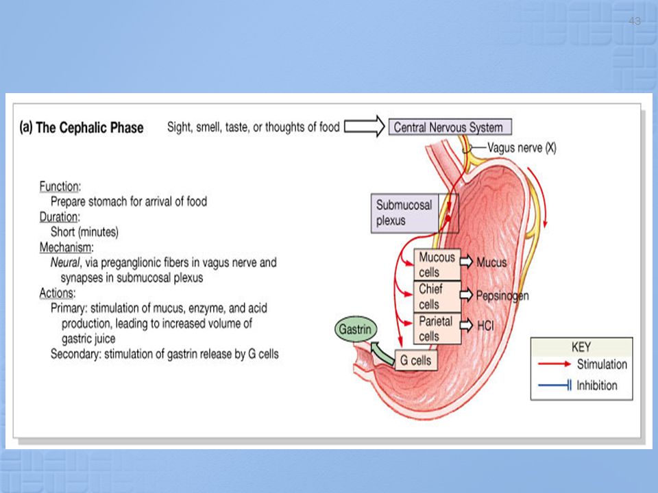
Phase 1: Cephalic reflex
The cephalic reflex phase of gastric secretion occurs before food enters the stomach It is triggered by the aroma, taste, sight, or though of food During this phase the brain gets the stomach ready for food

41
Phase 1: Cephalic reflex
Inputs from activated olfactory receptors and taste buds are relayed to the hypothalamus which in turn stimulates the vagal nuclei of the medulla oblongata, causing motor impulses to be transmitted via the vagus nerves to the parasympathetic nerve ganglia Eneteric ganglionic neurons in turn stimulate the stomach glands

42
Phase 1: Cephalic reflex
The enhanced secretory activity that results when we see or think of food is a conditioned reflex and occurs only when we like or want the food If we are depressed or have no appetite, this part of the cephalic reflex is suppressed

44
Phase 2: Gastric reflex Once food reaches the stomach, local neural and hormonal mechanisms initiate the gastric phase This phase provides about two-thirds of the gastric juice released The most important stimuli are distension, peptids, and low acidity

45
Phase 2: Gastric reflex Stomach distension activates stretch receptors and initiates both local (myentertic) reflexes and the long vagovagal reflexes In vagovagal reflex, impulses travel to the medulla and then back to the stomach via vagal fibers Both types of reflexes lead to acetylcholine (ACH) release, which in turn stimulates the output of more gastric juice by cells
 release, which in turn stimulates the output of more gastric juice by cells.")
46
Figure 24.15b

47
Phase 2: Gastric reflex Though neural influences initiated by stomach distension are important, the hormone gastrin probably plays a greater role in stimulating stomach gland secretion during the gastric phase Chemical stimuli provided by partially digested proteins (peptids)caffine (colas, coffee) and rising pH directly active gastrin secreting entoendocrine cells called G cells
caffine (colas, coffee) and rising pH directly active gastrin secreting entoendocrine cells called G cells.")
48
Phase 2: Gastric reflex Although gastrin also stimulates the release of enzymes, its main target is the HCL secreting parietal cells, which it prods to spew out even more HCL Highly acidic (pH below 2) gastric contents inhibit gastrin secretion
 gastric contents inhibit gastrin secretion.")
49
Phase 2: Gastric reflex When protein foods are in the stomach, the pH of the gastric contents generally rises because proteins act as buffers to tie up H+ The rise in pH stimulates gastrin and subsequently HCL release, which in turn provides the acidic conditions needed for protein digestion

50
Phase 2: Gastric reflex The more protein in the meal, the greater the amount of gastrin and HCL released As proteins are digested, the gastric contents gradually become more acidic, which again inhibits the gastrin secreting cells This negative feedback mechanism helps maintain optimal pH and working conditions for the gastric enzymes

51
Phase 2: Gastric reflex G cells are also activated by the neural reflexes already described Emotional upsets, fear, anxiety, or anything that triggers the fight-or-flight response inhibits gastric secretion because (during such times) the sympathetic division overrides parasympathetic controls of digestion
 the sympathetic division overrides parasympathetic controls of digestion.")
52
Phase 2: Gastric reflex The control of the HCL secreting parietal cells is unique and multifaceted Basically, HCL secretion is stimulated by three chemicals, all of which work through second-messenger systems Ach released by parasympathetic nerve fibers and gastrin secreted by G cells

53
Phase 2: Gastric reflex Ach released by para-sympathetic nerve fibers and gastrin secreted by G cells bring about their effects by increasing intercellular Ca++ levels

54
Histamine released by mucosal cells called histaminocytes acts through cyclic AMP (cAMP)
")
55
Phase 2: Gastric reflex As hydrogen ions are secreted, chloride ions (Cl-) are also pumped into the lumen to maintain an electrical balance in the stomach The Cl- is obtained from blood plasma, while the H+ appears to come from a breakdown of carbonic acid formed by the combination of carbon dioxide and water and within the parietal cells
 are also pumped into the lumen to maintain an electrical balance in the stomach. The Cl- is obtained from blood plasma, while the H+ appears to come from a breakdown of carbonic acid formed by the combination of carbon dioxide and water and within the parietal cells.")
56
CO2 + H2O H2CO3 H+ + HCO3- As H+ is pumped from the cell and HCO3- is ejected through the basal cell membrane into the capillary blood

57
Phase 2: Gastric reflex The result of ejection of the bicarbonate ion into the capillary blood is that blood draining from the stomach is more alkaline than the blood serving it The phenomenon is called the alkaline tide

58
Phase 3: Intestinal The intestinal phase of gastric secretion has two components One excitatory One inhibitory

59
Phase 3: Intestinal The excitatory aspect is set into motion as partially digested food begins to fill the initial part (duodenum) of the small intestine This stimulates intestinal mucosal cells to release a hormone that encourages the gastric glands to continue their secretory activity
 of the small intestine. This stimulates intestinal mucosal cells to release a hormone that encourages the gastric glands to continue their secretory activity.")
60
The effects of this hormone imitate those of gastrin, so it has been named intestinal (enteric) gastrin However, intestinal mechanisms stimulate gastrin secretion only briefly As the intestine distends with chyme containing large amounts of H+, fats, partially digested proteins, and irritating substances, the inhibitatory component is triggered in the form of the enterogastric reflex

61
The enterogastric reflex is actually a trio of reflexes that
Inhibit the vagal nuclei in the medulla Inhibit local reflexes Activate sympathetic fibers that cause the pyloric sphincter to tighten and prevent further food entry into the small intestine As a result, gastric secretory activity declines

62
These inhibitions on gastric activity product the small intestine to harm due to excessive acidity and match the small intestine’s processing abilities to the amount of chyme entering it at a given time

63
In addition, the factors just named trigger the release of several intestinal hormones collectively called enterogastrones which include Secretin Cholecystokinin (CCK) Vasoactive intestinal peptide (VIP) Gastric inhibitory peptide (GIP) All of these hormones inhibit gastric secretion when the stomach is very active
 Vasoactive intestinal peptide (VIP) Gastric inhibitory peptide (GIP) All of these hormones inhibit gastric secretion when the stomach is very active.")
64
Figure 24.15c

65
Regulation of Pancreatic Secretion
Secretion of pancreatic juice is regulated both by local hormones and by the parasympathetic nervous system

66
Regulation of Pancreas Secretion
Both hormones act on the pancreas, but secretin targets the duct cells, prompting their release of watery bicarbonate-rich pancreatic juice, Whereas CCK stimulates the acini to release enzyme-rich pancreatic juice Vagal stimulation causes release of pancreatic juice primarily during the cephalic and gastric phases of gastric secretion

67
Normally, the amount of HCL produced in the stomach is exactly balanced by the amount of bicarbonate (HCO3) actively secreted by the pancreas HCO3 is secreted into the pancreatic juice, and H+ enters the blood Consequently, the pH of venous blood returning to the heart remains relatively unchanged because alkaline blood draining from the stomach is neutralized by the acidic blood draining the pancreas

68
Regulation of Pancreatic Secretions
Secretin Acidity in intestines induces Secretin release Secretin releases pancreatic Sodium Bicarbonate (HCO3-) CCK Fats and proteins induce CCK release CCK releases pancreatic digestive enzymes GIP Fatty acids and sugar causes induce GIP release GIP induces insulin release
 CCK. Fats and proteins induce CCK release. CCK releases pancreatic digestive enzymes. GIP. Fatty acids and sugar causes induce GIP release. GIP induces insulin release.")
69
The Gallbladder Although the liver makes bile continuously bile does not usually enter the small intestine until the gallbladder contract The major stimulus for gallbladder contraction is the intestinal hormone cholecystokinin (CCK) CCK is released to the blood when acidic, fatty chyme enters the duodenum
 CCK is released to the blood when acidic, fatty chyme enters the duodenum.")
70
Besides causing the gallbladder to contract, CCk has two other important effects
It stimulates secreation of pancreatic juice It relaxes the hepatppancreatic sphincter so that bile and pancreatic juice can enter the duodenum Parasympathetic impulses delivered by the vagus nerves have a minor impact on stimulating gallbladder contraction

71
Defecation The rectum is usually empty, but when feces are forced into it by mass movements, stretching of the rectal walls initiates the defecation reflex

72
This is a spinal cord mediated reflex that causes the walls of the sigmoid colon and the rectum to contract and the anal sphincters to relax

73
Distension or stretch of the rectal walls triggers a depolarization of sensory (afferent) fibers which synapse with the spinal cord
 fibers which synapse with the spinal cord")
74
Parasympathetic motor (efferent) fibers, in turn, stimulate contraction of the rectal walls and relaxation of the internal anal sphincter
 fibers, in turn, stimulate contraction of the rectal walls and relaxation of the internal anal sphincter")
75
If it is convenient to defecate, voluntary signals stimulate the relaxation of the external anal sphincter

76
As feces are forced into the anal canal, impulses reach the brain allowing us to decide whether the external(voluntary) anal sphincter should remain open or closed If defection is delayed, the reflex contractions end within a few seconds and the walls relax With the next mass movement, the reflex is initiated again and again until one chooses to defecate

Presentasi serupa