Upload presentasi
Presentasi sedang didownload. Silahkan tunggu

1
OLEH SUDRAJAT FMIPA UNMUL 2009

2
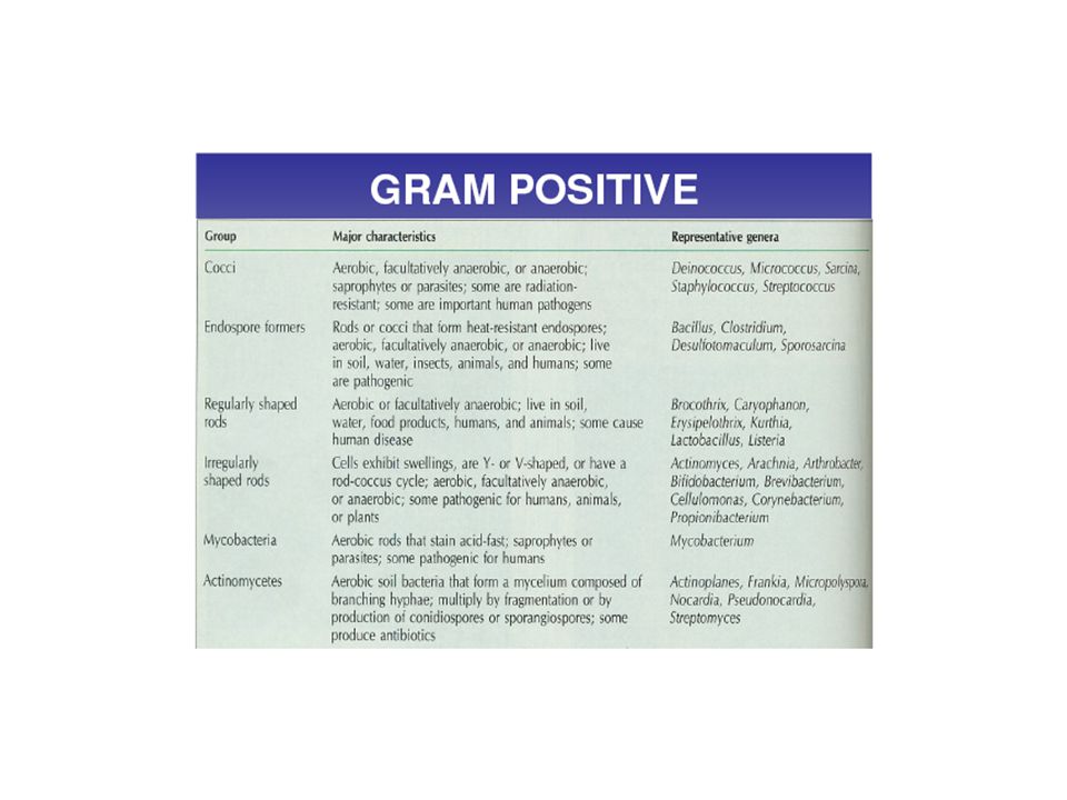
2 GRAM POSITIF Has a thick peptidoglycan layer –90% of the Gram-positive cell wall is comprised of peptidoglycan two types of teichoic acids

3
3 GRAM POSITIF 1) Lipoteichoic acid –on the surface, embedded in the peptidoglycan layer –linked to the cytoplasmic membrane
 Lipoteichoic acid –on the surface, embedded in the peptidoglycan layer –linked to the cytoplasmic membrane")
4
4 GRAM POSITIF 2) Wall teichoic acid –on the surface –linked to only the peptidoglycan layer
 Wall teichoic acid –on the surface –linked to only the peptidoglycan layer")
5
5 GRAM POSITIF

6
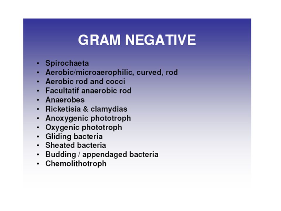
6 cell wall of Gram-negative bacteria is much thinner –comprised of only 20% peptidoglycan have two unique regions which surround the outer plasma membrane: –periplasmic space –lipopolysaccharide layer GRAM NEGATIF

7
7 a thin peptidoglycan layer an outer membrane attached to the peptidoglycan layer by lipoproteins

8
8 GRAM NEGATIF the outer membrane is made of protein, phospholipid and lipopolysaccharide –the lipid portion is embedded in the phospholipid –The lipid is toxic

9
9 GRAM NEGATIF –The cell wall has channels called Porins for the transport of low molecular weight substances

10
10 GRAM NEGATIF –periplasmic space between the cytoplasmic membrane and the cell wall –hydrolytic enzymes –antibiotic inactivating enzymes –transport proteins

11
11 Strong negative charge assists in: –evading phagocytosis –evade the complement system provides increased barrier to: –antibiotics, lysozymes, detergents GRAM NEGATIF

12
12 provides more attachment sites for: –virus –harmful substances more susceptible to mechanical breakage lipid A endotoxin is toxic to host GRAM NEGATIF

13
13 CELL WALL the cell wall is not a regulatory structure like the cell membrane though it is porous, it is not selectively permeable and will let anything pass that can fit through its gaps

14
Klasifikasi Bakteri Somewhat different: a clinical rapid ID is often important when trying to find causative agent of a disease Bergey’s manual: Manual is in lab for a reference when doing unknown Developed on 1940’s for grouping bacteria according to standard diagnostic lab techniques available at the time (such as Grams stain)
")
15
Klasifikasi Bakteri Gram +cocciGram - bacilli

16
Klasifikasi Bakteri Gram - SpirocheteGram + bacilli

17
Klasifikasi Bakteri The manual divides bacteria into 4 groups or divisions on the basis of their Cell Wall –1. Gram + (stain violet) –2. Gram - (destain, and are counterstained pink or reddish color) –3. Bacteria that lack a cell wall (mycoplasma) –4. organisms that have a cell wall lacking “peptidoglycan” (archaeobacteria – Now called “Archaea”
 –2. Gram - (destain, and are counterstained pink or reddish color) –3. Bacteria that lack a cell wall (mycoplasma) –4. organisms that have a cell wall lacking peptidoglycan (archaeobacteria – Now called Archaea .")
18
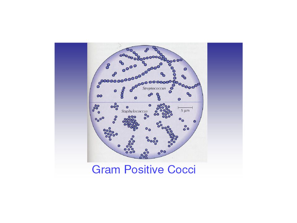
Microscopic Phenotypic Exam Gram stain –distinguishes between Gram + and Gram – bacteria –narrows the possibilities quickly Gram positive Gram negative

19
Microscopic Phenotypic Exam special stain allows for the distinction of microorganisms with unique characteristics capsule acid fast staining detects the waxy presence of Mycobacterium tuberculosis Capsule staining Acid fast staining of M. tuberculosis

20
Staining Organisms needed to allow us to see the organisms using light microscopy organisms are killed in the process Simple stains stain is applied and colours the organism e.g. methylene blue

21
Complex Stains stains may be combined which stain different structures different colours. e.g. giemsa stains malarial parasites nucleus red and cytoplasm blue stains may be applied in sequence with a step to remove stain in between. e.g. gram stain - a key stain in microbiology!!

22
The Gram Stain Developed by Christian Gram in the 19th Century He found that a stain could be washed out of some organisms much more easily than others Technique allows differentiation of many bacteria into 2 groups: gram positive and gram negative – corresponding to cell wall type. Continues to be used extensively and is important!

23
Method for Gram Stain Crystal violet – stains all the bacteria dark purple Iodine – binds to crystal violet and fixes it (acts as a mordant) Alcohol/Acetone washes out the stain from gram negative bacteria (Gram originally stopped here, so that organisms that stained purple were “positive” because they could be seen; subsequently the fourth step was added so that both the positive and the negative organisms could be seen.) Safranin stains the gram negative bacteria pink.
 Alcohol/Acetone washes out the stain from gram negative bacteria (Gram originally stopped here, so that organisms that stained purple were positive because they could be seen; subsequently the fourth step was added so that both the positive and the negative organisms could be seen.) Safranin stains the gram negative bacteria pink.")
24
Acid Fast Stain Some bacteria cannot be stained by the gram stain because of lipids in the cell walls. (e.g. Mycobacterium tuberculosis, the tuberculosis bacterium) These bacteria may be stained by an “acid fast method”. involves: - staining with a strong red stain (to “force” the stain in ) washing out the stain with a mixture of acid and alcohol restaining (“counterstaining”) with a blue or green stain. Acid Fast organisms are Red. These are sometimes called AFB (acid fast bacilli). Other organisms are the colour of the counter stain (blue or green).
 These bacteria may be stained by an acid fast method . involves: - staining with a strong red stain (to force the stain in ) washing out the stain with a mixture of acid and alcohol restaining ( counterstaining ) with a blue or green stain. Acid Fast organisms are Red. These are sometimes called AFB (acid fast bacilli). Other organisms are the colour of the counter stain (blue or green)..")
26
Ciri-ciri bakteri gram negatif a.Dinding sel tipis (10-15 nm) berlapis tiga (multi). b.Kandungan lipid tinggi : peptidoglikan (10% berat kering), tidak ada asam tekoat. c.Kerentanan terhadap penisilin kurang rentan. d.Pertumbuhan tidak begitu dihambat oleh zat warna dasar.
, tidak ada asam tekoat. c.Kerentanan terhadap penisilin kurang rentan. d.Pertumbuhan tidak begitu dihambat oleh zat warna dasar..")
27
Ciri-ciri bakteri gram negatif e.Persyaratan nutrisi → relatif sederhana. f.Resistensi terhadap gangguan fisik→ kurang resisten g.Kehilangan kompleks warna ungu kristal pada waktu dicuci alkohol → terwarnai pewarna tandingan safranin (sel tampak merah muda).
..")
30
Ciri – Ciri bakteri gram positif a.Struktur dinding sel tebal (15 – 80 nm) dan berlapis tunggal. b.Komposisi kimiawi : kandungan lipid rendah (1 - 4 %), peptidoglikan lapis tunggal (>50%), asam tekoat. c.Kerentanan terhadap penisilin→ lebih rentan (peka). d.Pertumbuhan dihambat oleh zat-zat warna dasar (misal ungu kristal)
, peptidoglikan lapis tunggal (>50%), asam tekoat. c.Kerentanan terhadap penisilin→ lebih rentan (peka). d.Pertumbuhan dihambat oleh zat-zat warna dasar (misal ungu kristal).")
31
Ciri - ciri bakteri gram positif e.Persyarataan nutrisi → relatif rumit pada banyak spesies. f.Resistensi terhadap gangguan fisik→ lebih resisten (tahan). g.Reaksi terhadap pewarna primer atau ungu kristal iodium → dapat menahan sampai akhir prosedur (sel tampak biru gelap/ungu).
. g.Reaksi terhadap pewarna primer atau ungu kristal iodium → dapat menahan sampai akhir prosedur (sel tampak biru gelap/ungu)..")
Presentasi serupa


>")


