Upload presentasi
Presentasi sedang didownload. Silahkan tunggu

1
SISTEM GASTROINTESTINAL Dr. ANNISA SITI ROHIMA

2
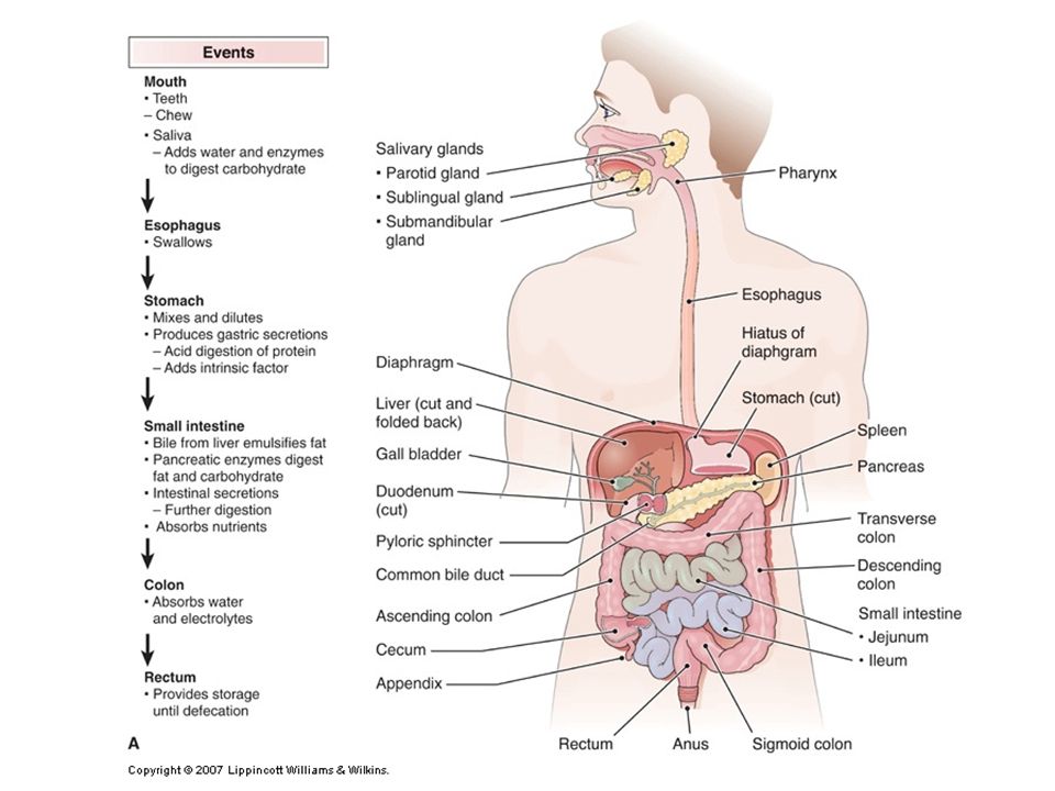
Traktus Digestivus/Gastrointestinalis -Traktus digestivus/gastrointestinalis bertugas untuk makan dan mengolah makanan, menyerap nutrisi dan membuang produk yang tidak terdigesti. - Traktus digestivus merupakan tabung panjang yang berawal dari cavum oris, esofagus, gaster, intestinum tenua, intestinum crassum/colon, rectum dan anus. -Kelenjar pencernaaan akan membantu mengolah dan menyerap nutrisi, yaitu glandula salivatorius, pankreas, hepar, dan vesica velea.

3
Struktur Sistem Organ Gastrointestinal Ingestion (mulut) Digesti (mulut, gaster, intestinum tenue) Organ asesorius (hepar, pankreas, vesica velea) Absorpsi (intestinum tenue et crassum) Eksresi (intestinum crassum)
 Digesti (mulut, gaster, intestinum tenue) Organ asesorius (hepar, pankreas, vesica velea) Absorpsi (intestinum tenue et crassum) Eksresi (intestinum crassum)")
5
Pengolahan Makanan Ingesti – Proses memasukkan makanan ke dalam tubuh Peristalsis – Gerakan fisik sepanjang traktus digestivus Digesti – Proses pengolahan makanan secara mekanik dan kimiawi Absorpsi – Perjalanan makanan dari traktus digestivus ke dalam tubuh Defekasi – Eliminasi bahan makanan yang tidak tercerna dalam tubuh

6
CRANIUM

7
Cranium: tampak lateral

9
Otot Kepala

10
Otot Mastikasi

11
1.M. masseter 2.M. buccinator ditembus oleh ductus glandula parotideus 3.M. pterigoideus medial 4.M. pterigoideus lateral

12
Cavum Oris Cavum oris: -Vestibulum oris -Cavum oris propria Ada: Arcus dentis/gigi Linguae/lidah

13
Dentes

14
Gigi/ Dentes Dentes decidui = gigi susu : 24 gigi Dentes permanentes = gigi tetap : 32 gigi Penyusun: Dens incicivus Dens caninus Dens premolaris Dens molaris I Dens molaris II Dens molaris III/serotinus

15
Mikroskopis Gigi Corona dentis, cervix dentis, radix dentis Enamel Dentinum Pulpa coronalis/ cavitas coronae Pulpa radicularis/ cavitas radicis dentis a, v, n

16
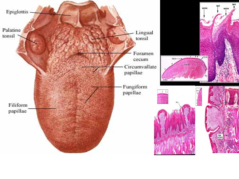
Lidah/Lingua Anatomi: radix, dorsum, apex linguae Musculus intrinsik: m.Transversus linguae m. Longitudinalis superior et inferor m. Verticalis linguae Papilla linguae: papilla valata, foliata, fungiformis dan piriformis

18
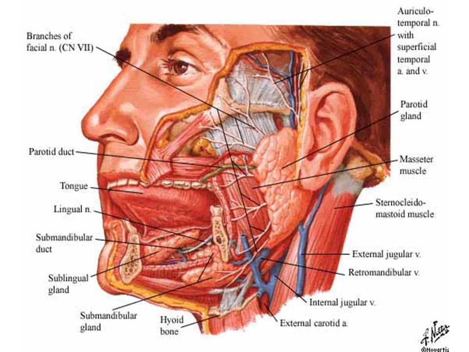
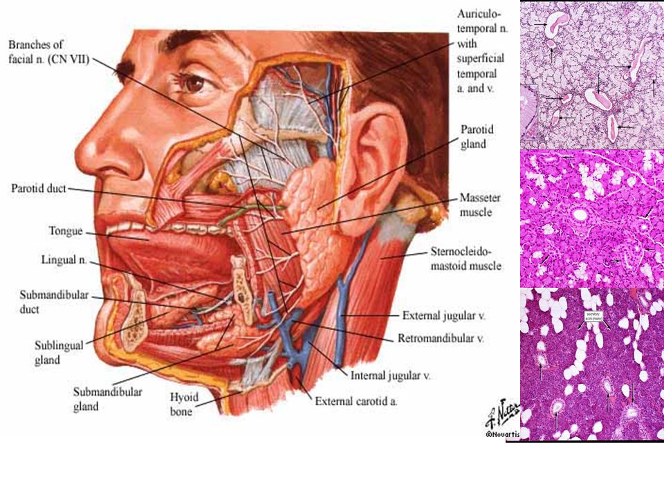
Glandula Salivariae Glandula salivares mayor: 1.Gl. Parotidea 2.Gl. Submandibularis 3.Gl. Sublingualis Glandula salivares minor: Gl. Platina

20
Traktus Digestivus/Gastrointestinalis Traktus penyusun gastrointestinal terdiri dari 4 lapisan yang berbeda: 1.Tunika mukosa 2.Tunika submukosa 3.Tunika muscularis 4.Tunika adventitia/serosa -Organ abdomen yang terletak retroperitoneal: duodenum, pancreas, colon ascendens, colon descendens, dan rectum

21
Faring Orofaring: Berbatasan dengan cavum oris melalui istmus faucium, meluas dari palatum molle hingga tepi atas epiglotis

22
Faring

23
Musculus Faring M. constrictor pharingis superior M. constrictor pharingis medius M. constrictor pharingis inferius M. palatoglossus M. salpingopharingeus M. stylopharingeus

24
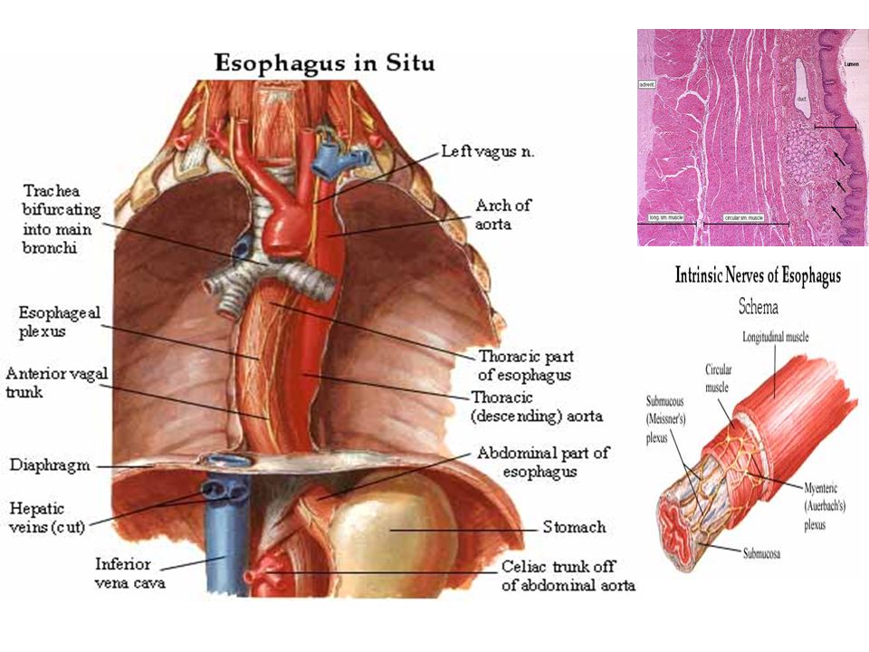
Esofagus Esofagus merupakan pipa berotot dengan panjang 25 cm Terbentang antara faring hingga gaster/ventriculus Bermuara pada cardia ventriculi setinggi cartilago costalis VII dan vertebra T10/11 Distal terdapat pleksus esofagealis

26
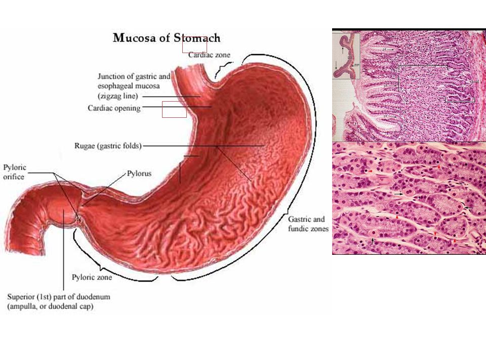
Gaster/Ventriculus/Lambung Bagian: cardiac, fundus, corpus, pilorus Cardiac merupakan muara esofagus Pilorus memiliki m. sfingter pylori untuk mengatur pengosongan lambung ke duodenum Lengkung: curvatura major et minor

27
NOTE: - The stomach sits in the upper left quadrant of the abdomen. It can be divided into 4 parts: the cardia, the fundus, the body or corpus, and the pylorus. - The lesser curvature of the stomach is connected to the liver via the hepatogastric ligament, which comprise the lesser omentum with the hepatoduodenal ligament. On the other side, the greater curvature is connected to the greater omentum of the abdomen. Note the other surrounding structures. - The venous drainage of the lesser curvature involves the left and right gastric veins, which anastomose as the coronary vein. The greater curvature is drained by short gastric veins into the anastomoses of the left and right gastro-omental veins. They all drain into the hepatic portal vein, hepatic veins, and inferior vena cava.

28
- The stomach is supplied by the arteries branching off the celiac trunk. - There are three major branches of the celiac trunk: - 1) left gastric artery – supplies the lesser curvature and anastomoses with the right gastric artery - 2) splenic artery – supplies the spleen, giving off the left gastro-omental artery which supplies the greater curvature and anastomoses with the right gastro- omental artery - 3) common hepatic artery – supplies the liver with the hepatic artery proper. The right gastric and right gastro-omental arteries both branch off the hepatic artery proper. In addition, it also gives off the gastroduodenal artery to supply the duodenum, pancreas, and greater curvature. - In short, the stomach is supplied by the right and left gastric arteries at the lesser curvature and the right and left gastro-omental arteries at the greater curvature. - The lesser curvature is drained by the coronary vein, while the greater curvature is drained by the right and left gastro- omental veins.
 left gastric artery – supplies the lesser curvature and anastomoses with the right gastric artery - 2) splenic artery – supplies the spleen, giving off the left gastro-omental artery which supplies the greater curvature and anastomoses with the right gastro- omental artery - 3) common hepatic artery – supplies the liver with the hepatic artery proper. The right gastric and right gastro-omental arteries both branch off the hepatic artery proper. In addition, it also gives off the gastroduodenal artery to supply the duodenum, pancreas, and greater curvature. - In short, the stomach is supplied by the right and left gastric arteries at the lesser curvature and the right and left gastro-omental arteries at the greater curvature. - The lesser curvature is drained by the coronary vein, while the greater curvature is drained by the right and left gastro- omental veins..")
30
Duodenum

31
Merupakan bagian terpendek dari intestinum tenue, berbentuk huruf C, berhubungan dengan pylorus dan jejunum Anatomis: pars superior, pars decendens, pars horizontalis, dan pars ascendens Terdapat muara ductus pancreaticus dan ductus choledocus/biliaris

32
Jejunum dan Ileum

33
- Once again, the 4 layers of the GI tract are shown – mucosa, submucosa, muscular layers, and serosa. - The jejunum and ileum are attached to the posterior abdominal wall via mesentery. Within the mesentery are arcades and straight arteries. Jejunal arteries are shorter than ileal arteries. In addition, the jejunum mucosa has many more circular folds than the ileum, showing that the jejunum absorbs most of the nutrients. - Histologically, the jejunum and ileum are very similar. Note once again the numerous villi. Extending into the lamina propria from the mucosa are intestinal glands, better known as intestinal crypts or crypts of Lieberkuhn. - REMEMBER: Only the esophagus and duodenum have submucosal glands.

34
Jejunum dan Ileum Berfungsi sebagai penyerapan

35
- The ileum ends in the right lower quadrant of the abdomen and connects to the cecum, which then leads into the ascending colon. - The ileocecal region is supplied by the ileocolic artery, which branches off the superior mesenteric artery. The ileocolic artery gives off a colic branch which supplies beginning of the ascending colon, and an ileal branch that supplies the end of the ileum. - Note the abrupt transition in the epithelial lining from the small intestinal (S) villi to the glandular form of large intestine (L). The ileocecal valve contains considerably thickened muscularis propria (M) with some lymphoid tissue (Ly). - Note the appendix and appendicular artery shown here. We look in more detail in two slides. CAECUM
 villi to the glandular form of large intestine (L). The ileocecal valve contains considerably thickened muscularis propria (M) with some lymphoid tissue (Ly). - Note the appendix and appendicular artery shown here. We look in more detail in two slides. CAECUM.")
36
Intestinum Crassum/Colon

37
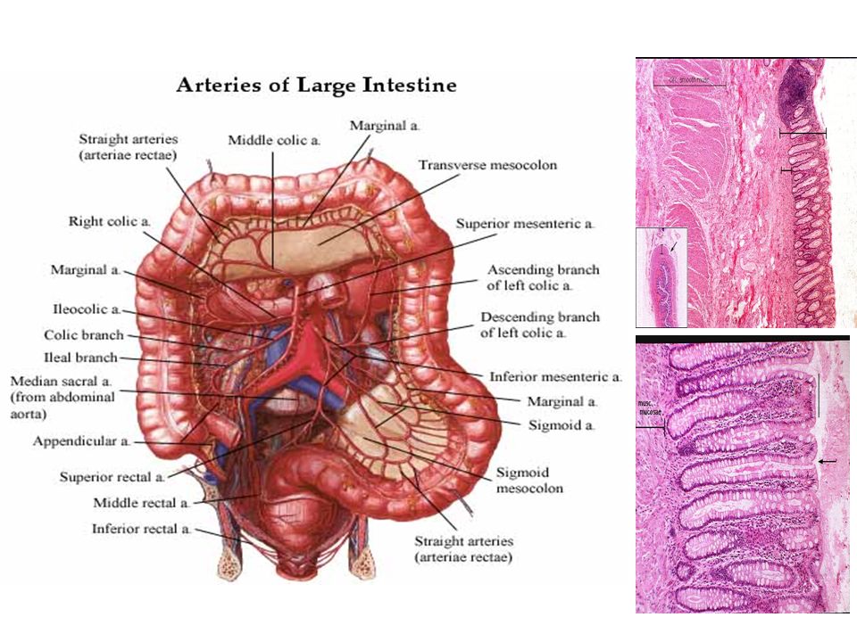
Intestinum crassum/ Colon Anatomis: colon ascendens, transversum, decendens, sigmoideum Rectum merupakan bagian akhir dari colon

39
ANUS Memiliki 2 sfingter: Sfingter ani interna Sfingter ani eksterna

Presentasi serupa