Upload presentasi
Presentasi sedang didownload. Silahkan tunggu

1
Pendahuluan Biologi Molekuler

2
DNA DOGMA CENTRAL RNA Protein
I’m betting that an intimate understanding of cell cycle regulation and apoptosis is key to understanding the process of carcinogenesis. If nothing else, the main idea emphasized here is that the coupling between the cell cycle and apoptosis – how these processes are coordinated – is key. This talk will focus on the links and controls of the onset of the cell cycle and apoptosis. Mathematical Biosciences Institute (Ohio State Univ), 2 October 2003
, 2 October")
3
Dogma central Biologi Molekuler
DNA Sequence (splited by genes) RNA Asam amino protein fenotip Adapted from
 RNA. Asam amino. protein. fenotip. Adapted from")
4
DNA Gula Fosfat Basa (A,T, C or G)
DNA adalah komponen yang tersusun dari dari molekul-molekul yang disebut nukleotid Masing-masing nukleotid mengandung fosfat, gula dan basa nitrogen. Ada empat basa: Adenine (A), Guanine (G), Cytosine (C), and Thymine (T)
, Guanine (G), Cytosine (C), and Thymine (T)")
5
DNA: structure Ikatan gula-fosfat pada nukleotid merupakan backbone dari ikatan pada DNA Empat basa dari DNA dibentuk sepanjang “backbone” disebut dengan DNA sequence. Dua DNA saling berikatan di antara pasangan basa Dua ikatan basa yang mungkin yaitu: A-T, C-G. Dua untai DNA mempunyai formasi struktur double helixTwo. Source of diagram:

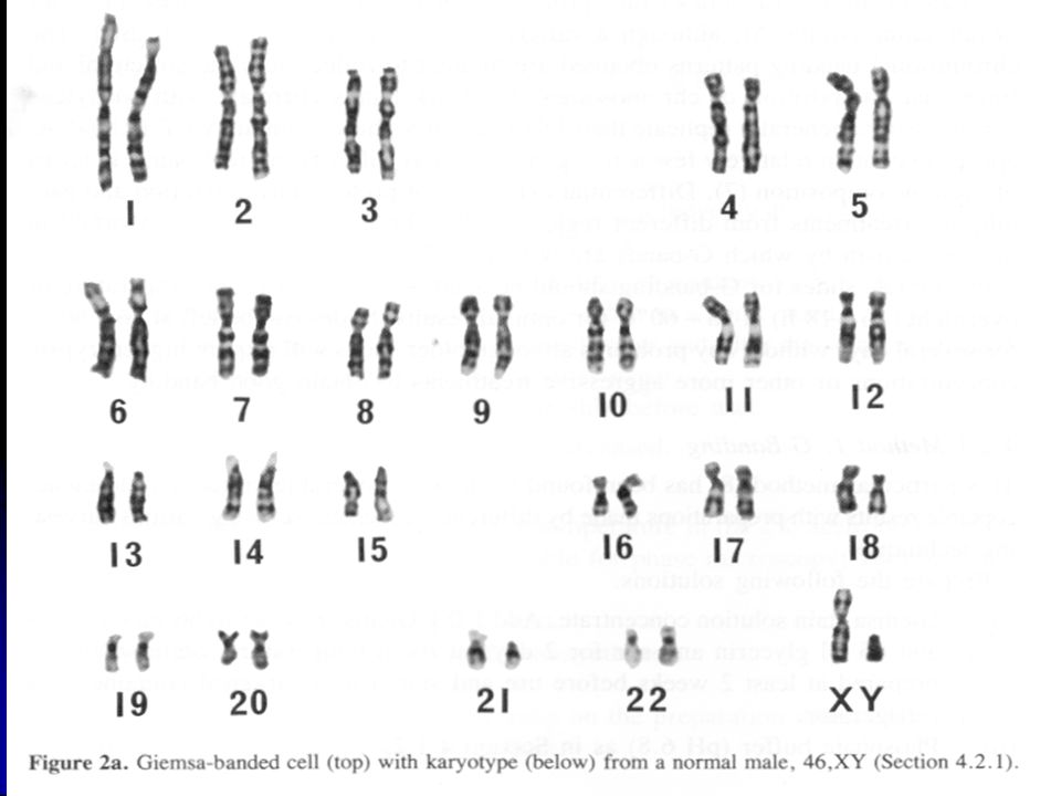
6
DNA: tersusun dalam kromosom
Each chromosome is essentially a package for a very long, continuous DNA double strand. Lodish et al. Molecular Biology of the Cell (5th ed.). W.H. Freeman & Co., 2003.
. W.H. Freeman & Co.,")
7
DNA: di-splid oleh gen-gen
promoter Exon 1 Intron 1 Exon 2 Intron 2 Exon 3 gene Gen merupakan bagian dari DNA yang membawa informasi untuk membentuk protein. 2-3% dari DNA manusia adalah gen, gen yang tidak aktif (rest) disebut junk DNA Promotor terlelak di bagian awal dari komponen gen. Promotor aktif saat gen akan bekerja. Pada banyak gen euryotic, gen adalahsekuen DNA yang mempunyai kode, yang juga disebut sebagai exon. Bagian yang tidak membawa kode genetik disebut sebagai intron.
 disebut junk DNA. Promotor terlelak di bagian awal dari komponen gen. Promotor aktif saat gen akan bekerja. Pada banyak gen euryotic, gen adalahsekuen DNA yang mempunyai kode, yang juga disebut sebagai exon. Bagian yang tidak membawa kode genetik disebut sebagai intron.")
8
RNA RNA (ribonucleic acid) adalah intermediet antara DNA dan protein.
RNA merupakan single strand dari asam nukleat. Basa nitrogen T (Thymine) pada RNA terganti oleh U (Uracil) Tidak seperti DNA, yang terlokasi di inti, RNA juga dapat ditemukan di sitoplasma. Pada inti, kode gen ditranskripsikan pada RNA. Selanjutnya RNA akan keluar dari inti ke nukleus dalam sitoplasma, dimana RNA ditranslasi menjadi asam amino. Source of diagram:
 pada RNA terganti oleh U (Uracil) Tidak seperti DNA, yang terlokasi di inti, RNA juga dapat ditemukan di sitoplasma. Pada inti, kode gen ditranskripsikan pada RNA. Selanjutnya RNA akan keluar dari inti ke nukleus dalam sitoplasma, dimana RNA ditranslasi menjadi asam amino. Source of diagram:")
9
Another view of central dogma
Gen diekspresikan pada 3 step: 1) Transkripsi: Sintesis RNA 2) Splicing: penghilangan intron dari RNA 3) Translasi: Sintesis Protein
 Transkripsi: Sintesis RNA. 2) Splicing: penghilangan intron dari RNA. 3) Translasi: Sintesis Protein.")
10
Transkripsi Animation
Transkripsi diinisiasi oleh kompleks dari faktor=faktor transkripsi yang berikatan dengan promotor. An enzyme, RNA polymerase II, travels along the gradually unzipped DNA template and polymerizes nucleotides into an RNA. The sequence of nucleotides on DNA template determines the sequence on RNA by following the rule of base-pair complementarity, i.e., A – U, T- A, C – G, G – C. Transcription continues until entire gene is copied to RNA. Animation Source of diagram:

11
Splicing pre mRNA Exon 1 Intron 1 Exon 2 Intron 2 Exon 3 mature mRNA

12
Translation (1) By translation, the nucleotide sequence on mRNA
determines the amino acid sequence by genetic code. Genetic code: three base pairs of RNA (called a codon) determine one amino acid based on a fixed table. Translation always starts at AUG (start codon), and ends with any of UAA, UAG, or UGA (stop codon)
 determine one amino acid based on a fixed table. Translation always starts at AUG (start codon), and ends with any of UAA, UAG, or UGA (stop codon)")
13
Translation (2) Transfer RNAs (tRNAs): small RNA molecules. Most of the tRNAs function as carriers of amino acids and participate in protein synthesis. For example, the tRNA with the anticodon CGG corresponds with the codon GCC and attaches alanine amino acid onto the peptide chain. Ribosome: a complex of protein and rRNA Animation Source of diagram:
: small RNA molecules. Most of the tRNAs function as carriers of amino acids and participate in protein synthesis. For example, the tRNA with the anticodon CGG corresponds with the codon GCC and attaches alanine amino acid onto the peptide chain. Ribosome: a complex of protein and rRNA. Animation. Source of diagram:")
14
Summary Central dogma of molecular biology Three components
DNA RNA Protein Three steps transcription splicing translation

16
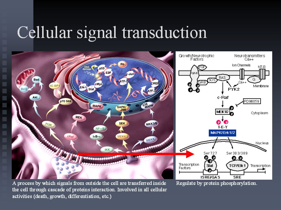
Cell – cell communication

18
Proto-oncogenes encode components of growth factor signal transduction pathways
Components shown in yellow are known proto-oncogenes

19
The VEGF family and its receptors
VEGF-A VEGF-B PlGF VEGF-A VEGF-C VEGF-D VEGF receptor-2 VEGF receptor-1 VEGF receptor-3 P– – P – P P– – P P– P– – P P– – P P– – P Two types of VEGF receptors have been identified. VEGF receptor-1 (Flt-1) and VEGF receptor-2 (KDR/Flk-1) bind VEGF with high affinity. Both VEGF receptor-1 and VEGF receptor-2 have seven immunoglobulin (Ig)-like domains in the extracellular domain (ECD). VEGF receptor-2 has the highest binding affinity for VEGF165.1 Another member of the receptor family with seven Ig-like domains in the ECD is VEGF receptor-3 (Flt-4). Though not a receptor for VEGF, it binds VEGF-C and -D. In the embryo, VEGF receptor-3 is expressed in angioblasts and venules. Later on in development, it becomes restricted to lymphatic endothelium, suggesting that it has a role in the regulation of lymphangiogenesis.1 Ferrara N, Gerber HP, LeCouter J. The biology of VEGF and its receptors. Nat Med 2003;9:669–76. Migration, permeability, DNA synthesis, survival Angiogenesis Lymphangiogenesis Adapted from Ferrara N. Nat Med 2003;9:669–76
 and VEGF receptor-2 (KDR/Flk-1) bind VEGF with high affinity. Both VEGF receptor-1 and VEGF receptor-2 have seven immunoglobulin (Ig)-like domains in the extracellular domain (ECD). VEGF receptor-2 has the highest binding affinity for VEGF Another member of the receptor family with seven Ig-like domains in the ECD is VEGF receptor-3 (Flt-4). Though not a receptor for VEGF, it binds VEGF-C and -D. In the embryo, VEGF receptor-3 is expressed in angioblasts and venules. Later on in development, it becomes restricted to lymphatic endothelium, suggesting that it has a role in the regulation of lymphangiogenesis.1. Ferrara N, Gerber HP, LeCouter J. The biology of VEGF and its receptors. Nat Med 2003;9:669–76. Migration, permeability, DNA synthesis, survival. Angiogenesis. Lymphangiogenesis. Adapted from Ferrara N. Nat Med 2003;9:669–76.")
20
VEGF signal transduction and its effects
Permeability VEGF-C VEGF VEGF-D VEGF receptor-1 VEGF receptor-2 Cation channel VEGF receptor-3 P– – P – P P– – P P– P– – P Ca2+ P– PLC – P P– – P IP3 PLC Calcium release DAG PLC Evidence suggests that VEGF receptor-1 and VEGF receptor-2 have differing signal transduction properties and may mediate different functions. VEGF receptor-2 undergoes strong ligand-dependent tyrosine phosphorylation in intact endothelial cells and mediates mitogenesis and chemotaxis in response to VEGF, whereas VEGF receptor-1 produces weak or undetectable responses. It has been suggested that VEGF receptor-1 is not a signalling receptor but a ‘decoy’ receptor, negatively regulating the activity of VEGF on the vascular endothelium by sequestering and rendering this ligand less available to VEGF receptor-2. Conversely, later studies show that VEGF receptor-1 can interact with signal-transducing proteins and generate a mitogenic signal. More recent studies suggest that VEGF receptor-1’s main role is as a ligand-binding receptor, rather than a signal-transducing receptor.1 VEGF is also thought to have a role in the control of HSC survival and repopulation by means of an internal autocrine loop, i.e. the ligand interacts with its receptors within the cell.2 Mouse studies suggest, in contrast to their action on endothelial cells, that both VEGF receptor-1 and VEGF receptor-2 have roles in HSC development and survival.2 These observations could have implications for the toxicity of intracellular receptors blockers, e.g. tyrosine kinase inhibitors such as PTK787, which may interfere with the autocrine loop. Externally activating agents such as bevacizumab would not be expected to have these effects. Stimulation of signalling through VEGF receptor-2 on endothelial cells by VEGF binding results in the activation of a number of signalling pathways, including the MAPK and ras pathways. This results in cellular effects including endothelial cell proliferation, survival and migration, as well as increased vascular permeability.3 Ferrara N. Role of vascular endothelial growth factor in the regulation of angiogenesis. Kidney Int 1999;56:794–814. Gerber H-P, Ferrara N. The role of VEGF in normal and neoplastic hematopoiesis. J Mol Med 2003;81:20–31. Shibuya M. Structure and function of VEGF/VEGF-receptor system involved in angiogenesis. Cell Struct Funct 2001;26:25–35. DAG P13K Protein kinase C Raf-1 MAPK SAPK/ JNK Protein kinase B Proliferation, migration Permeability Apoptosis Survival Proliferation Migration VEGF binding to VEGF receptor-2 activates a signalling cascade resulting in cellular effects Shibuya M. Cell Struct Funct 2001;26:25–35

21
Agents targeting the VEGF pathway
Antibodies inhibiting VEGF receptors Soluble VEGF receptors (VEGF-TRAP) Antibodies inhibiting VEGF (e.g. bevacizumab) Permeability VEGF Cation channel VEGF receptor-2 Small-molecules inhibiting VEGF receptors (TKIs) (e.g. PTK-787) P– – P – P – P P– P– – P P– Over expression of VEGF by tumour cells can be targeted by: antibodies against VEGF antibodies against VEGF receptors soluble VEGF receptors that bind circulating VEGF small molecule inhibitors of VEGF receptors catalytic RNA molecules (ribozymes), which cleave VEGF receptor mRNA. P– P– – P – P Migration, permeability, DNA synthesis, survival Ribozymes (Angiozyme) Angiogenesis Lymphangiogenesis
 Antibodies inhibiting VEGF (e.g. bevacizumab) Permeability. VEGF. Cation channel. VEGF receptor-2. Small-molecules inhibiting VEGF receptors (TKIs) (e.g. PTK-787) P– – P. – P. – P. P– P– – P. P– Over expression of VEGF by tumour cells can be targeted by: antibodies against VEGF. antibodies against VEGF receptors. soluble VEGF receptors that bind circulating VEGF. small molecule inhibitors of VEGF receptors. catalytic RNA molecules (ribozymes), which cleave VEGF receptor mRNA. P– P– – P. – P. Migration, permeability, DNA synthesis, survival. Ribozymes. (Angiozyme) Angiogenesis. Lymphangiogenesis.")
22
Signal Transduction from Receptor to Nucleus Via RAS p21
GDP Sos Grb2 P GTP Ras Growth factor P120-GAP Neurofibromin 14-3-3 1 2 3 Inactive Raf Transcription factors etc. DNA synthesis Nucleus MEK ERK1 ? Active Raf P13-K Rac and Rho pathway Morphological change

23
Regulators : proliferation, differentiation, apoptosis, repair
Transcriptome RNA Genome : Genes : Cell cycle Differentiatio Apoptosis Repair Metabolism etc Proteome Protein: cyclin, CDK,CDKI GF : GM-CSF, FGF Bcl-2, p53, caspase Gadd, enzym repair dll

Presentasi serupa


, YANG TERSUSUN DARI SENYAWA NUKLEOTID. CONTOH SENYAWA ASAM.>")


