Upload presentasi
Presentasi sedang didownload. Silahkan tunggu

1
Program Studi Ilmu Keperawatan Unitri Malang
Human anatomy SKELETAL SYSTEM dr. Dea Amanda Program Studi Ilmu Keperawatan Unitri Malang

2
Anatomi.. : Ilmu yg mempelajari ttg struktur tubuh
Tubuh sistem organ organ jaringan sel Sistem organ: muskuloskeletal, GIT, urinary, reproductive, respiratory system, cardiovascular Organ: cardia, gaster, ren, uterus, prostat, pulmo, ureter

3
Fisiologi.. : Ilmu yg mempelajari ttg proses tubuh N Cardiovaskuler:
Memompa darah ke seluruh tubuh u/ myalurkan O2 & nutrisi GIT: Mencerna makanan u/ diserap & bahan bakar tubuh Muskuloskeletal: bergerak

4
Anatomical Position & Terminology
Posisi Anatomis: the Body is erect/upright Legs together Feet are parallel, directed forwards, flat on the floor Arms at the sides of the body Palms turned forward, with fingers pointing downwards

5
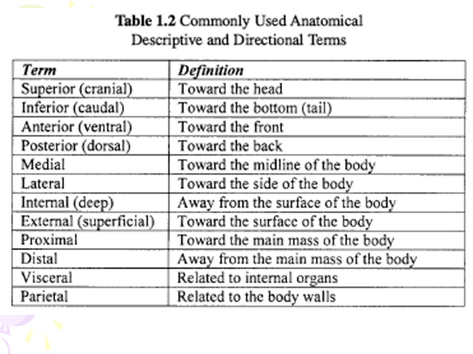
Istilah2.. (=terminology)
Superior (=cranial) vs Inferior (=caudal) Anterior (=ventral) vs Posterior (=dorsal) Medial vs Lateral Internal (=deep) vs Eksternal (=superfisial) Proksimal vs Distal
 vs Inferior (=caudal) Anterior (=ventral) vs Posterior (=dorsal) Medial vs Lateral. Internal (=deep) vs Eksternal (=superfisial) Proksimal vs Distal.")
8
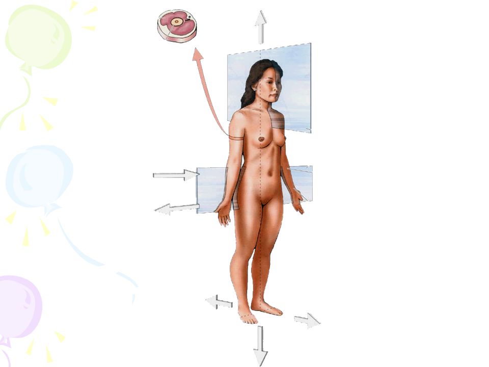
Bidang2 Tubuh.. Sagital (kiri-kanan); mid-sagital: (pas tengah)
Koronal (depan-belakang) Transversal (superior-inferior)
 Transversal (superior-inferior)")
9
Skeletal System Structure: Bones Cartilage Joints ∑ tulang:206

10
Skeletal System.. (2) Function Support :penopang tubuh
Protection :melindungi organ2 vital Movement :bergerak saat kontraksi otot Hematopoiesis :terlindung dlm sutul, produksi sel darah Mineral storage

11
Klasifikasi tulang.. (brdsrkn bentuk)
Tulang panjang: ada beda panjang>lebar (femur, tibia, humerus) Tulang pendek: spt kubus (carpal, tarsal) Tulang pipih: proteksi (skull, ribs, scapula) Tulang ireguler: bentuk aneh (vertebrae, face)
 Tulang pendek: spt kubus (carpal, tarsal) Tulang pipih: proteksi (skull, ribs, scapula) Tulang ireguler: bentuk aneh (vertebrae, face)")
12
General structures of bone
Bone substance compact bone spongy bone ※flat bones 2 layers compact bone (outer plate & inner plate); spongy bone in the middle
; spongy bone in the middle.")
13
Long Bone Structure.. Diaphysis: di tgh Epiphysis: Medullary cavity:
di ujung prox&dist Tdd tulang spongious dikelilingi tulang kompak Medullary cavity: rongga dlm diafisis; dilapisi endosteum; Berisi: yellow bone marrow.

14
Long Bone Structure.. Metaphysis: Epiphyseal plate:
Daerah dmn diafis bergabung dg epifis Epiphyseal plate: Mitosis>>, u/ elongasi Saat pertumb slesai epiphyseal line Articular cartilage: Kartilago melapisi tiap epiphysis.

15
Periosteum: Outer or fibrous layer
Inner layer vascular & contains osteoblasts Tempat menempelnya tendon

16
Long Bone Structure.. Figure 6.3

17
Histologi..

18
Histologi.. (2) Central Canal: circular channelcontains blood, lymphatic vessels, & nerves. (Concentric )Lamellae: calcified matrix surrounding a vertically oriented blood vessel. Lacuna: a small hollow space, contains osteocytes. Canaliculus: a small channelconnects lacunae to each other, & to the central canal.
Lamellae: calcified matrix surrounding a vertically oriented blood vessel. Lacuna: a small hollow space, contains osteocytes. Canaliculus: a small channelconnects lacunae to each other, & to the central canal.")
19
Bone Formation.. Terdapat bbrp tipe sel tulang, a.l:
Sel osteogenik :sel progenitor (=stem sel) Osteoblast sel2 tulang mghasilkan osteoid Calcium & mineral lain mengeraskan osteoid Osteosit :osteoblast trapped inside osteoid develop jd osteosit Osteoklas :bone digestion - sel pghancur tulang demineralisasi sel tulang
 Osteoblast. sel2 tulang mghasilkan osteoid. Calcium & mineral lain mengeraskan osteoid. Osteosit :osteoblast trapped inside osteoid develop jd osteosit. Osteoklas :bone digestion - sel pghancur tulang demineralisasi sel tulang.")
20
Bone Cell Types

21
Bone Formation.. = osifikasi Dimulai pd mgg ke-4 dlm kandungan
Trdpt 2 cara: Endochondral – mll thp kartilago Kondrosit (sel tlg rwn) hipertrofi kalsifikasi Intramembranous –lgsg terbentuk sbg tulang keras
 hipertrofi kalsifikasi. Intramembranous –lgsg terbentuk sbg tulang keras.")
22
Long Bone Formation and Growth..

23
Bone Growth in Length.. Epiphyseal plate
Cartilage cells in this plate divide rapidly. Zone of proliferating cartilage. Between ages 18-25, the epiphyseal plates close. Cartilage cells in the plate stop dividing and bone replaces the cartilage. Growth in length stops at age 25.

24
Bone Growth in Width..

25
Bone Remodeling.. Saat massa tulang optimum telah tercapai remodelling mjd proses utama pd tulang Fungsi: (1) memperbaiki kerusakan mikro tulang (2) mempertahankan kekuatan tulang (3) men-supply Ca dari tulang untuk mempertahankan kadar Ca serum.
 memperbaiki kerusakan mikro tulang. (2) mempertahankan kekuatan tulang. (3) men-supply Ca dari tulang untuk mempertahankan kadar Ca serum.")
26
Bone Remodeling.. Hasil akhir proses remodelling: tulang yg diresorpsi diganti dg tulang baru Pembentukan & resorpsi tulang proses normal, & continuous

27
SKELETON (Kerangka Tulang)
Axial : Skull 22 Auditory ossicles 6 Hyoid bone 1 Vertebral column 26 Ribs and sternum 25 ---- 80 Appendicular : Upper extremity 64 Lower extremity 62 Total 206 Axial Skeleton: merupakan tulang axis dari tubuh cranium, vertebrae, costae, sternum Appendicular Skeleton: berbungan dengan penyusun tubuh : extremitas atas, bawah, & pelvis

28
The Axial & Appendicular Skeleton
Figure 5.6 Slide 5.20b Copyright © 2003 Pearson Education, Inc. publishing as Benjamin Cummings

29
Joints.. (=articulation)
: tempat pertemuan 2 tulang / lebih Klasifikasi: Synarthrosis (immovable) Amphiarthrosis (slightly movable) Diarthrosis (freely movable)
 Amphiarthrosis (slightly movable) Diarthrosis (freely movable)")
30
Synarthrosis.. ≠ dpt bergerak
Tulang yg 1 dg yg lain dihubungkan dg jar. fibrosa Contoh: Sutura pd tlg2 tengkorak Tibia-fibula (distal)
")
31
Amphiarthrosis.. dpt sedikit bergerak
Ujung2 tulangnya dibungkus tulang rawan hyalin Contoh: Symphisis pubis Intervertebral joints

32
Diarthrosis.. (=sendi synovial)
Sendi yg dpt digerakkan dg bebas Memiliki rongga sendi Ujung2 tulang dilapisi tulang rawan hyalin Contoh: Lutut Siku

33
Pergerakan Sendi.. Fleksi vs Ekstensi Abduksi vs Adduksi Rotasi
Pronasi vs Supinasi

34
To Be Continued..

Presentasi serupa


, FICS, MPd>")


>")


