Upload presentasi
Presentasi sedang didownload. Silahkan tunggu

1
Wanita 34 tahun dengan Rubella
LAPSUS 2 POLIKLINIK 24/12/2013 Wanita 34 tahun dengan Rubella Anugerah Pembimbing: Dr. Rivan D, Sp.S, M.Kes. 1

2
IDENTITAS PENDERITA Nama : Ny. S P Umur : 34 tahun
Jenis kelamin : Wanita Pekerjaan : Karyawati Agama : Islam Alamat : Ngasem, Colomadu Tgl Pemx : 17 Desember 2013 pk WIB No. RM :

3
I. ANAMNESIS Keluhan Utama Nyeri kepala sebelah kanan
Keluhan Yang Menyertai Kelemahan anggota gerak sebelah kanan

4
Riwayat Penyakit Sekarang
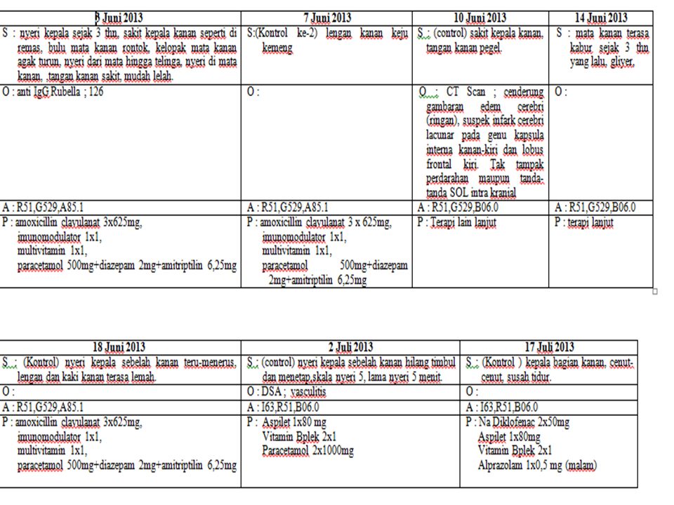
Keluhan utama : nyeri kepala Penderita merasa nyeri kepala sejak 3 tahun ini. Nyeri kepala sebelah kanan, dari mata kanan sampai kepala bagian belakang. Nyeri seperti di remas-remas, hingga mengeluarkan air mata. Nyeri di rasakan terus menerus. Nyeri tidak disertai mual atau pun muntah. Juga disertai pandangannya semakin lama semakin kabur, terutama mata sebelah kanan. Pada jarak 1 meter penderita bisa melihat jari tangan, tetapi kabur. Bulu mata penderita juga rontok. Disertai pandangan dobel dalam 6 bulan ini. Tidak pernah bengkak atau benjolan di daerah telinga atau tempat lainnya.

5
Riwayat Penyakit Sekarang
Sudah pernah di rawat di RS Swasta 2 kali. Pertama pada tahun 2010 : menderita sakit kepala biasa. Tahun 2011 : keluhan yang sama. CT Scan : terkena stroke Derajat nyeri dari awal sampai sekarang, dirasakan sama.

6
Riwayat Penyakit Sekarang
Bulan April 2012 : periksa Laboratorium Toxo dan Rubella Ig G Rubella (+) Berobat di poliklinik RSDM mulai bulan Juni 2013, nyeri kepala sebelah kanan, saat itu juga disertai badannya sebelah kanan gringgingan, dan kekuatan anggota gerak sebelah kanan sedikit menurun. Mudah lelah. Kemudian disarankan rawat inap. Menjalani pemeriksaan DSA hasil pemeriksaan disimpulkan vaskulitis cerebral.
 Berobat di poliklinik RSDM mulai bulan Juni 2013, nyeri kepala sebelah kanan, saat itu juga disertai badannya sebelah kanan gringgingan, dan kekuatan anggota gerak sebelah kanan sedikit menurun. Mudah lelah. Kemudian disarankan rawat inap. Menjalani pemeriksaan DSA hasil pemeriksaan disimpulkan vaskulitis cerebral.")
7
Riwayat Penyakit Dahulu
Riwayat sakit sendi : (-) Riw. pembesaran kelenjar : (-) Riwayat sakit tenggorokan : (-) Riw. ISPA : (-) Riwayat perdarahan : (-) Riw. demam lama : (-) Riwayat mondok : (+) Riw. Hipertensi : (-) Riwayat cedera kepala : (-)
 Riw. pembesaran kelenjar : (-) Riwayat sakit tenggorokan : (-) Riw. ISPA : (-) Riwayat perdarahan : (-) Riw. demam lama : (-) Riwayat mondok : (+) Riw. Hipertensi : (-) Riwayat cedera kepala : (-)")
8
RIWAYAT PENYAKIT KELUARGA
Riwayat penyakit yang sama : (-)
")
9
Keadaan sosial ekonomi
Pasien : seorang karyawati, (bekerja di pabrik pewarna celana jeans dan sepatu) status janda, satu anak. Berobat dengan fasilitas Jamkesmas. Riwayat Kebiasaan dan Gizi Riwayat olahraga : jarang Memelihara hewan : (-) Keadaan gizi : kesan cukup
 status janda, satu anak. Berobat dengan fasilitas Jamkesmas. Riwayat Kebiasaan dan Gizi. Riwayat olahraga : jarang. Memelihara hewan : (-) Keadaan gizi : kesan cukup.")
10
II. PEMERIKSAAN FISIK Status Interna : Kesan Umum : Kompos mentis
gizi kesan cukup Tanda Vital : TD : 120/70 mmHg RR : 20 x/mnt N : 80 x/mn T : 36,5°C VAS : 4 Kepala : dbn Leher : KGB (-), JVP ≠ ↑ Ketiak&lipatan paha : pembesaran KGB (-)
, JVP ≠ ↑ Ketiak&lipatan paha : pembesaran KGB (-)")
11
Status Interna : Jantung : I : iktus cordis tidak tampak : P : iktus kordis tidak kuat angkat : P : kesan batas jantung tidak melebar : A : BJ I-II reguler, bising(-) Paru : I : pengembangan simetris : P : fremitus raba kiri = kanan : P : sonor / sonor : A : vesikuler ( +/+ ), suara tambahan ( - / - )
 Paru : I : pengembangan simetris. : P : fremitus raba kiri = kanan. : P : sonor / sonor. : A : vesikuler ( +/+ ), suara tambahan ( - / - )")
12
Status Interna : Status Psikiatri :
Abdomen : I : cembung, vena kolateral (-) : P : supel, hepar & lien tidak teraba : P : tympani : A : bising usus (+) Status Psikiatri : Emosi : dbn Proses berpikir : dbn Kecerdasan : dbn Perhatian : dbn
 : P : supel, hepar & lien tidak teraba. : P : tympani. : A : bising usus (+) Status Psikiatri : Emosi : dbn. Proses berpikir : dbn. Kecerdasan : dbn. Perhatian : dbn.")
13
Status Neurologis Kesan Umum & Fungsi luhur Kepala : dbn Kesadaran : komposmentis, E4 V5 M6 Cara berbicara : dbn Fungsi psikosensorik : agnosia sensorik (-) agnosia motorik (-) Fungsi psikomotorik :dbn
 agnosia motorik (-) Fungsi psikomotorik :dbn")
14
Status Neurologis Meningeal Sign Kaku kuduk : (-) Tanda Laseque : (-)
Tanda Kernig : (-) Tanda Brudzinski I : (-) Tanda Brudzinski II : (-) Tanda Brudzinski III : (-) Tanda Brudzinski IV : (-)
 Tanda Brudzinski I : (-) Tanda Brudzinski II : (-) Tanda Brudzinski III : (-) Tanda Brudzinski IV : (-)")
15
Status Neurologis Kolumna Vertebralis Kelainan bentuk : (-)
Nyeri tekan / ketok : (-) Tanda Patrick : (-) Tanda Kontrapatrick : (-) Gerak V. Cervikal : dbn Gerak tubuh : dbn
 Tanda Patrick : (-) Tanda Kontrapatrick : (-) Gerak V. Cervikal : dbn. Gerak tubuh : dbn.")
16
Status Neurologis (Saraf Otak )
Nervus I (Olfaktorius) Anosmia : - / - Parosmia : / - Halusinasi : - / - Nervus II (Optikus) Kanan Kiri Visus : >3/ >3/60 Kacamata : (-) (-) Lapang Pandang : dbn dbn Warna : dbn dbn Funduskopi : dbn dbn
 Anosmia : - / - Parosmia : - / - Halusinasi : - / - Nervus II (Optikus) Kanan Kiri. Visus : >3/60 >3/60. Kacamata : (-) (-) Lapang Pandang : dbn dbn Warna : dbn dbn. Funduskopi : dbn dbn.")
17
Status Neurologis (Saraf Otak)
Nervus III, IV, VI Kanan Kiri - Celah mata : Simetris Posisi bola mata Ditengah Gerak bola mata dbn Pupil : ukuran Bentuk 3 mm Bulat R. cahaya langsung (+) R. cahaya tak langsung Konvergensi Akomodasi
 R. cahaya tak langsung. Konvergensi. Akomodasi.")
18
Status Neurologis (Saraf Otak )
Nervus V Kanan Kiri Sensorik I : ↓ dbn Sensorik II : ↓ dbn Sensorik III : ↓ dbn Otot kunyah : dbn dbn Refleks masseter : dbn dbn Refleks kornea :

19
Status Neurologis (Saraf Otak )
Nervus VII Saat diam Saat gerak kanan kiri kanan kiri Otot dahi : simetris simetris Tinggi alis : simetris simetris Sudut mata : simetris simetris Sudut mulut : simetris simetris Lipatan nasolabial : simetris simetris Memejamkan mata : simetris simetris Meringis : simetris simetris Sekresi air mata : dbn Pengecap lidah : dbn Hiperakusis : / -

20
Status Neurologis (Saraf Otak )
Nervus VIII Kanan kiri Pendengaran : dbn dbn Vertigo : (-) (-) Nistagmus : (-) (-)
 (-) Nistagmus : (-) (-)")
21
Status Neurologis (Saraf Otak )
Nervus IX dan Nervus X Kanan Kiri Refleks muntah : dbn dbn Pengecapan : dbn dbn Posisi uvula : ditengah Arkus faring : dbn Menelan : dbn Bersuara : dbn Fenomena Vernet Rideau: simetri

22
Status Neurologis (Saraf Otak )
Nervus XI Kanan Kiri Bentuk otot : dbn dbn Mengangkat bahu : dbn dbn Berpaling : dbn dbn

23
Status Neurologis (Saraf Otak )
Nervus XII Kanan Kiri Atrofi lidah : (-) (-) Kekuatan : dbn dbn Fasikulasi : (-) (-) Posisi diam : di tengah Posisi dijulurkan : ditengah Gerak spontan : dbn
 (-) Kekuatan : dbn dbn. Fasikulasi : (-) (-) Posisi diam : di tengah. Posisi dijulurkan : ditengah. Gerak spontan : dbn.")
24
Status Neurologis (Sistem Koordinasi)
Kanan Kiri Gerakan abnormal : (-) (-) Uji jari-jari tangan : dbn dbn Uji jari hidung : dbn dbn Uji pronasi dan supinasi : dbn dbn Uji hidung-jari-hidung : dbn dbn Uji tumit lutut : dbn dbn Cara berjalan : dbn Uji Romberg : (-)
 (-) Uji jari-jari tangan : dbn dbn. Uji jari hidung : dbn dbn. Uji pronasi dan supinasi : dbn dbn. Uji hidung-jari-hidung : dbn dbn. Uji tumit lutut : dbn dbn. Cara berjalan : dbn. Uji Romberg : (-)")
25
Status Neurologis (Sistem Sensorik )
Lengan Tungkai Kanan Kiri kanan kiri Rasa eksteroseptif Rasa nyeri superfisial : dbn dbn dbn dbn Rasa suhu : dbn dbn dbn dbn Rasa raba ringan : dbn dbn dbn dbn Rasa proprioseptif Rasa getar : dbn dbn dbn dbn Rasa tekan : dbn dbn dbn dbn Rasa nyeri tekan : dbn dbn dbn dbn Rasa gerak dan posisi : dbn dbn dbn dbn

26
Status Neurologis (Sistem Sensorik )
Rasa kortikal kanan kiri Stereognosis : dbn dbn Barognosis : dbn dbn Pengenalan 2 titik (atas) : dbn dbn (bawah) : dbn dbn
 : dbn dbn (bawah) : dbn dbn.")
27
Status Neurologis (Sistem Otonom)
Miksi : dbn Defekasi : dbn Salivasi : dbn Sekresi keringat : dbn

28
Status Neurologis Sistem Motorik & Refleks
Ekstremitas Superior Lengan Atas bawah tangan Kanan kiri kanan kiri kanan kiri Pertumbuhan : N N N N N N Tonus : N N N N N N Kekuatan Fleksi : Ekstensi : Reflek fisiologis Bisep : / Trisep : / Reflek patologis Hoffman : ( / ) Tromner : ( / )
 Tromner : (+ / -)")
29
Status Neurologis Sistem Motorik & Refleks
Ekstremitas Inferior Tungkai atas bawah kaki Kanan kiri kanan kiri kanan kiri Pertumbuhan : N N N N N N Tonus : N N N N N N Kekuatan Fleksi : Ekstensi : Klonus Lutut : (-) Kaki : (-)
 Kaki : (-)")
30
Status Neurologis Sistem Motorik & Refleks
Refleks Fisiologis & Patologis Kanan Kiri Refleks Patella : Refleks Achilles : Reflkes Babinski : (-) (-) Refleks Chaddock : (-) (-) Refleks Openheim : (-) (-) Refleks Gordon : (-) (-) Refleks Schaeffer : (-) (-) Refleks dinding perut : (-) (-)
 (-) Refleks Chaddock : (-) (-) Refleks Openheim : (-) (-) Refleks Gordon : (-) (-) Refleks Schaeffer : (-) (-) Refleks dinding perut : (-) (-)")
31
Status Neurologis Sistem Motorik & Refleks
Refleks Primitif Refleks memegang : (-) Refleks snout : (-) Refleks menghisap : (-) Refleks palmo-mental : (-)
 Refleks snout : (-) Refleks menghisap : (-) Refleks palmo-mental : (-)")
32
III. PEMERIKSAAN PENUNJANG
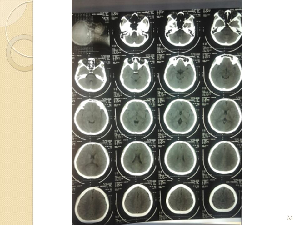
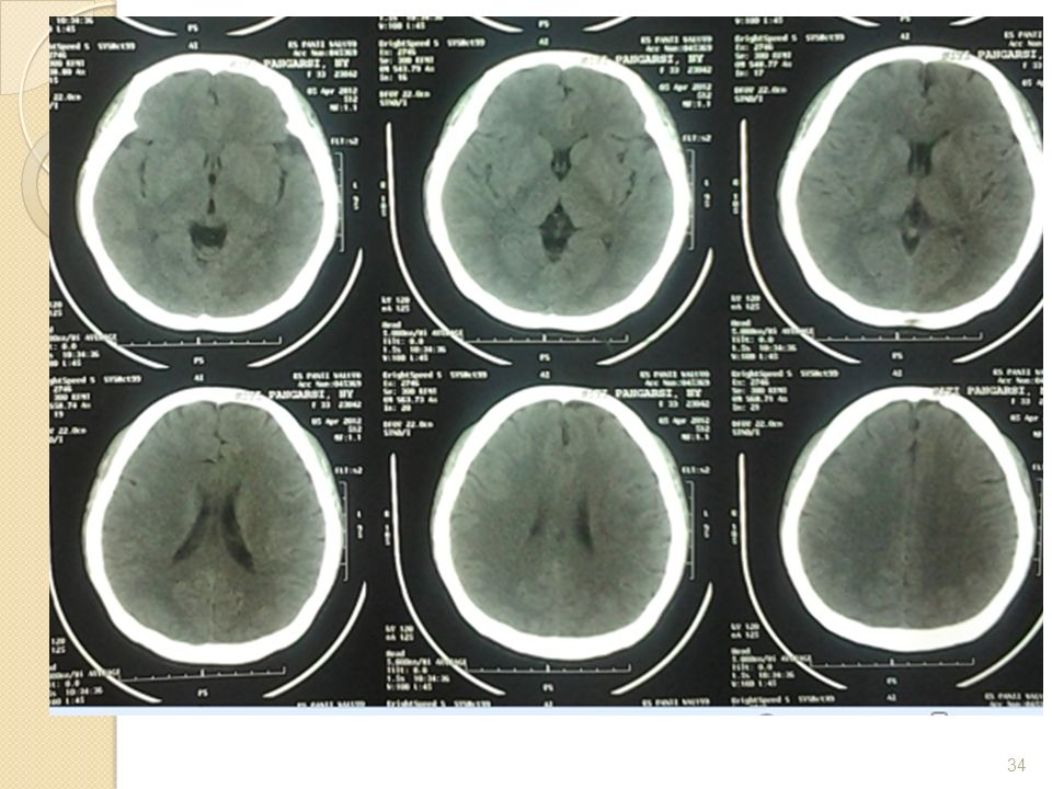
Pemeriksaan CT Scan Kepala polos (5/4 2012) Tampak lesi hipodens pada genu capsula interna kanan kiri dan lobus frontalis kiri Sulkus kortikalis region basal fisura serta sisterna sedikit menyempit System ventrikel normal Tak tampak midline shifting Tak tampak kelainan pada batang otak dan cerebellum KESAN :- Gambaran oedem cerebri ringan - Suspek infark cerebri lakunar pada genu kapsula interna kanan kiri dan lobus frontalis kiri - Tak tampak tanda tanda SOL intrakranial
 Tampak lesi hipodens pada genu capsula interna kanan kiri dan lobus frontalis kiri. Sulkus kortikalis region basal fisura serta sisterna sedikit menyempit. System ventrikel normal. Tak tampak midline shifting. Tak tampak kelainan pada batang otak dan cerebellum. KESAN :- Gambaran oedem cerebri ringan. - Suspek infark cerebri lakunar pada genu kapsula interna kanan kiri dan lobus frontalis kiri. - Tak tampak tanda tanda SOL intrakranial.")
36
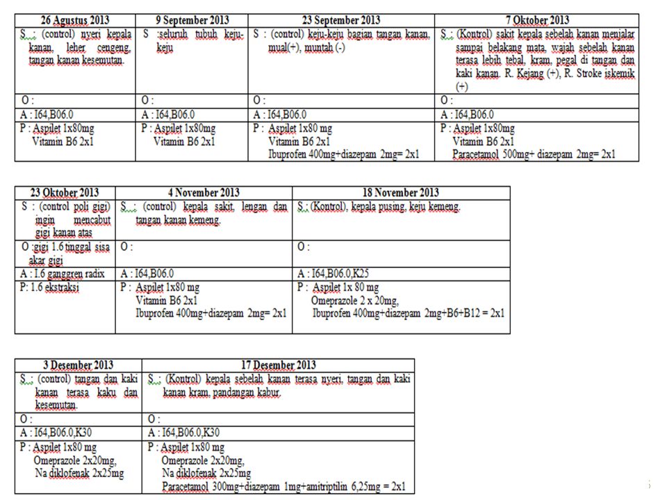
DSA tgl 26 Juni 2013 RCCA injeksi : normal, tidak ada stenosis di bifurkasio RICA injeksi : tampak normal aliran kontras sampai fase vena LCCA injeksi : normal, ada stenosis di bifurkasio LICA injeksi : tampak normal aliran kontras sampai fase vena. Tampak hipervaskularisasi arteriole dan venule yang menyokong gambaran inflamasi. Kesimpulan : Cerebral DSA menyokong gambaran vaskulitis cerebral Saran : Terapi di lanjutkan

38
IV. RESUME Nyeri kepala 3 tahun
Anamnesis Nyeri kepala 3 tahun Nyeri kepala bagian kanan. Nyeri seperti di remas remas, kadang kadang sampai mengeluarkan air mata. Nyeri di rasakan terus menerus. Derajat nyeri dari awal penderita merasakan nyeri sampai sekarang, dirasakan sama. Pandangan double Kelemahan anggota gerak kanan Gringgingan pada badan sebelah kanan Tidak ada riwayat trauma

39
IV. RESUME Pemeriksaan Fisik Status interna : dalam batas normal
Status neurologis : Kesadaran : GCS E4V5M6, compos mentis Fungsi sensoris : hemihipestesi dextra Fungsi motorik : hemiparese dextra Reflek fisiologis : meningkat pada lengan kanan Reflek patologis : Tromer (+) pada bagian kanan Nervi craniales : N V. Hemihipestesi dextra
 pada bagian kanan. Nervi craniales : N V. Hemihipestesi dextra.")
40
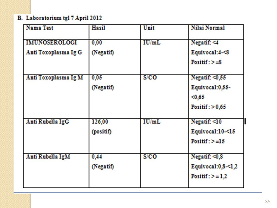
IV. RESUME Pemeriksaan Penunjang CT Scan : Suspek infark cerebri lakunar pada genu kapsula interna kanan kiri dan lobus frontalis kiri Laboratorium : Anti Rubella IgG (+) ,00 IU/ml DSA : Vaskulitis Cerebral
 126,00 IU/ml. DSA : Vaskulitis Cerebral.")
41
V. DIAGNOSIS Diagnosis Neurologis
Diagnosis klinis : Chepalgia kronik, diplopia, hemiparese dekstra, hemihipestesi dextra. Diagnosis topis : Subcortex sinistra Diagnosis etiologi: Rubella dengan Vaskulitis Cerebral Diagnosis lain : Stroke Infark

42
VI. PENATALAKSANAAN A. Pengobatan medikamentosa Aspilet 80 mg 1x1
B comp 2x1 Na diklofenak 2x25 mg Parasetamol 300mg,amitriptilin 6,25mg,diazepam 1mg caps 2x1

43
VII. KONSULTASI / RAWAT BERSAMA
Bagian Mata tgl 17 Des 2013 Jawaban : Visus : OD: 6/6 OS : 6/6 Papil oedem : tidak ditemukan Ptosis ringan

46
TERIMA KASIH

48
Rubella Disease Rare in children
20-50% of infections are asymptomatic Prodrome Rare in children Adolescents/adults: low grade fever, malaise, lymphadenopathy, upper respiratory symptoms lasts1- 5 days Rash Maculopapular Begins on face and head Usually persists 3 days Other symptoms Lymphadenopathy: postauricular, posterior cervical, and suboccipital Conjunctivitis

49
Congenital Rubella Syndrome
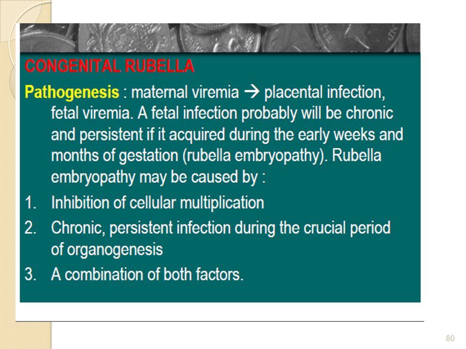
Infection in pregnancy, most dangerous <12 weeks gestation May lead to fetal death or premature delivery Hearing impairment, cataracts, heart defects, microcephaly, developmental delay, bone alterations, liver and spleen damage Organ specificity generally related to stage of gestational infection

50
Three day measles; German measles or
Rubella is an infectious disease transmitted by the rubella virus. The signs of the disease are: swelling of lymph nodes in the neck and a skin rash which first appears on the face and then rapidly spreads to other parts of the body. The complications of rubella include encephalitis and joint pain. Infection with rubella during pregnancy can cause serious damage to the child, including cataracts, deafness, heart defects and mental retardation. There is no drug therapy for rubella. However, the disease can be prevented by immunization.

51
The disease is caused by a virus that is spread through the air or by close contact. It can also be transmitted to a fetus by a mother with an active infection. The disease is usually mild and may even go unnoticed. Children may have few symptoms, but adults may experience a prodrome (warning symptom) of a fever, headache, malaise, runny nose, and inflamed eyes that lasts from 1 to 5 days before the rash appears. A person can transmit the disease from 1 week before the onset of the rash until 1 week after the rash disappears. The disease is not as contagious as rubeola (measles), therefore many people are not infected during childhood. Lifelong immunity to the disease follows infection. Epidemics may occur at about 6- to 9-year intervals. The risk factors are the unimmunized individuals. The disease is potentially serious because of the ability to produce defects in a developing fetus if the mother is infected during early pregnancy. As many as 10 to 15% of women in their childbearing years are susceptible to infection. Congenital rubella syndrome occurs in 25% or more of infants born to women who acquired rubella during the first trimester ofpregnancy. Defects are rare if the infection occurs after the 20th week of pregnancy. One or more defects may occur in an infected fetus and include deafness, cataracts,microcephaaly,mental retardation,congenital heart defects and other defects. A miscarriage or stillbirth may occur. Risk factors include lack of immunization and exposure to an active case of rubella.

52
Rubella syndrome, or congenital rubella, is a group of physical abnormalities that have developed in an infant as a result of maternal infection and subsequent fetal infection with rubella virus. It is characterized by rash at birth, low birth weight, small head size, heart abnormalities, visual problems and bulging fontanelle.

53
VACCINE INFORMATION The MMR vaccine is a "3-in-1" vaccine that protects against measles, mumps, and rubella. Although single antigen (individual) vaccines have been developed for each component of the MMR, they are not readily available and usually used only for very specific situations. An example of such a situation would be if an outbreak of either measles, mumps, or rubella was occurring in a specific community and public health officials deemed it necessary to immunize infants 6 to 12 months old. Single antigen vaccines might be used because they pose less risk to children younger than the recommended age of 12 months for the MMR. For children 12 months or older and adults, the risks of giving the single antigen vaccine are presumed to be the same as giving the MMR. The first shot is recommended at months. Because the first shot may not provide adequate lifetime immunity to some individuals, a second MMR is recommended prior to school entry at 4-6 years or prior to entry into junior high at years. Some states require a second MMR at kindergarten entry

54
Rubella Pathogenesis Respiratory transmission of virus
Replication in nasopharynx and regional lymph nodes Viremia 5-7 days after exposure with spread to tissues Placenta and fetus infected during viremia

55
Rubella Clinical Features
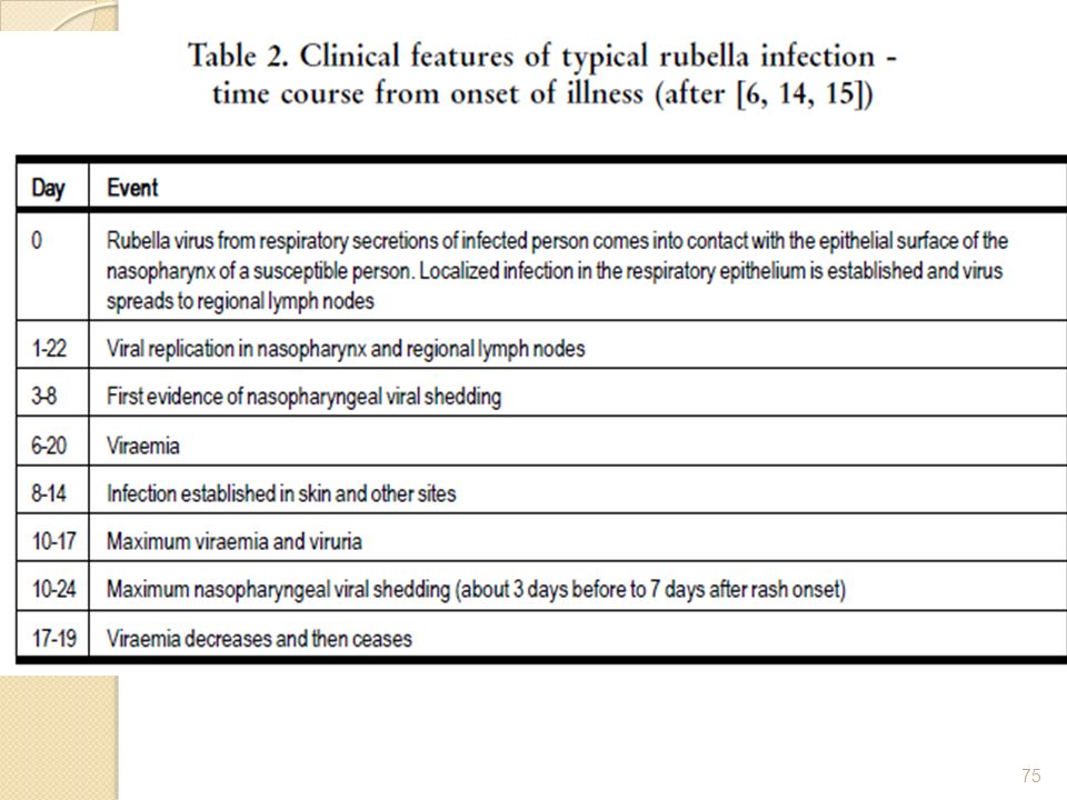
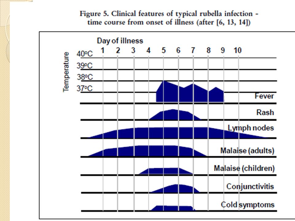
Incubation period 14 days (range days) Prodrome of low grade fever Lymphadenopathy in second week Maculopapular rash days after exposure
 Prodrome of low grade fever. Lymphadenopathy in second week. Maculopapular rash days after exposure.")
56
Rubella Complications
Arthralgia or arthritis children adult female Thrombocytopenic purpura Encephalitis Neuritis Orchitis rare up to 70% 1/3000 cases 1/5,000+ cases

57
Rubella Laboratory Diagnosis
Isolation of rubella virus from clinical specimen (e.g., nasopharynx, urine) Significant rise in rubella IgG by any standard serologic assay (e.g., enzyme immunoassay) Positive serologic test for rubella IgM antibody
 Significant rise in rubella IgG by any standard serologic assay (e.g., enzyme immunoassay) Positive serologic test for rubella IgM antibody.")
58
Rubella Epidemiology Reservoir Human
Transmission Respiratory Subclinical cases may transmit Temporal pattern Peak in late winter and spring Communicability 7 days before to 5-7 days after rash onset Infants with CRS may shed virus for a year or more

59
Rubella Vaccine Composition Live virus (RA 27/3 strain)
Efficacy 95% (Range, 90%-97%) Duration of Immunity Lifelong Schedule 1 Dose Should be administered with measles and mumps as MMR
 Duration of Immunity Lifelong. Schedule 1 Dose. Should be administered with measles and mumps as MMR.")
60
Eye pain on lateral and upward eye movement (a particularly troublesome complaint)
Conjunctivitis Sore throat Headache General body aches Low-grade fever Chills Anorexia Nausea Tender lymphadenopathy (particularly posterior auricular and suboccipital lymph nodes) Forchheimer sign (an enanthem observed in 20% of patients with rubella during the prodromal period; can be present in some patients during the initial phase of the exanthem; consists of pinpoint or larger petechiae that usually occur on the soft palate)
 Forchheimer sign (an enanthem observed in 20% of patients with rubella during the prodromal period; can be present in some patients during the initial phase of the exanthem; consists of pinpoint or larger petechiae that usually occur on the soft palate)")
61
Temperature Fever is usually not higher than 38.5°C (101.5°F). Lymph nodes Enlarged posterior auricular and suboccipital lymph nodes are usually found on physical examination. Mouth The Forchheimer sign may still be present on the soft palate.

62
Salt and pepper retinopathy
Courtesy Courtesy: Jonathan Trobe, M.D. - University of Michigan Kellogg Eye Center Content Providers(s): CDC Creation Date: 1976
: CDC Creation Date:")
63
DIFFERENTIALS Drug Eruptions Erythema Infectiosum (Fifth Disease) Measles, Rubeola Mucocutaneous Lymph Node Syndrome (Kawasaki Disease) Roseola Infantum Scarlet Fever
 Measles, Rubeola Mucocutaneous Lymph Node Syndrome (Kawasaki Disease) Roseola Infantum Scarlet Fever.")
64
Lab Studies If the diagnosis is in doubt, a rising titer of immunoglobulin M (IgM) antibody over a 2-week period indicates a recent infection. A WBC count, if performed, may be lower than normal, as in many viral infections, with increased percentages in the lymphocyte count. In those very rare cases where encephalitis is present, lymphocytes are present in the cerebrospinal fluid (CSF).
.")
65
Rubella virus can be isolated from the nasopharynx, the blood, the urine, and CSF.
Now, newer tests with a higher specificity (eg, latex agglutination, fluorescence immunoassay, passive hemagglutination, hemolysis in gel, enzyme immunoassay tests) are available. From a public health standpoint, attempting to confirm a rubella infection in a pregnant woman or a newborn or an infant is important.
 are available. From a public health standpoint, attempting to confirm a rubella infection in a pregnant woman or a newborn or an infant is important.")
66
A pregnant woman exposed to rubella should be tested immediately for a rubella-specific antibody. The presence of rubella-specific immunoglobulin G (IgG) is evidence that the patient is immune. If the test result is negative, repeat the test again in 3-4 weeks, and also repeat the test on the first specimen. If an antibody is present in the second test and not in the first test, an infection has occurred.

67
An infant with CRS will show the IgG antibody from the mother, which disappears in a few months and an elevated IgM antibody level because of antibody production by the infant. The presence of the IgM antibody usually indicates recent infection because IgM does not cross the placenta from the mother as does IgG

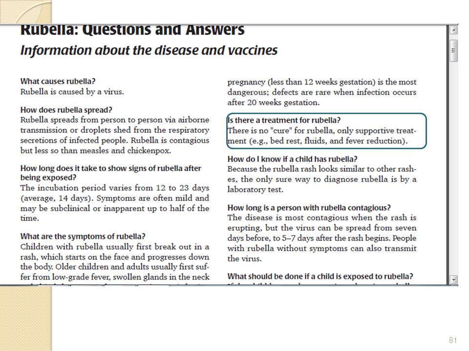
68
TREATMENT Medical Care: No specific treatment of rubella exists.
The disease is usually self-limited. Rest and oral fluids are appropriate. Individuals may remain contagious for 7 days after the onset of the rash, and they should be appropriately isolated from work, school, or other public settings.

69
Medication No specific medication is available for rubella, except that given for symptomatic relief.

72
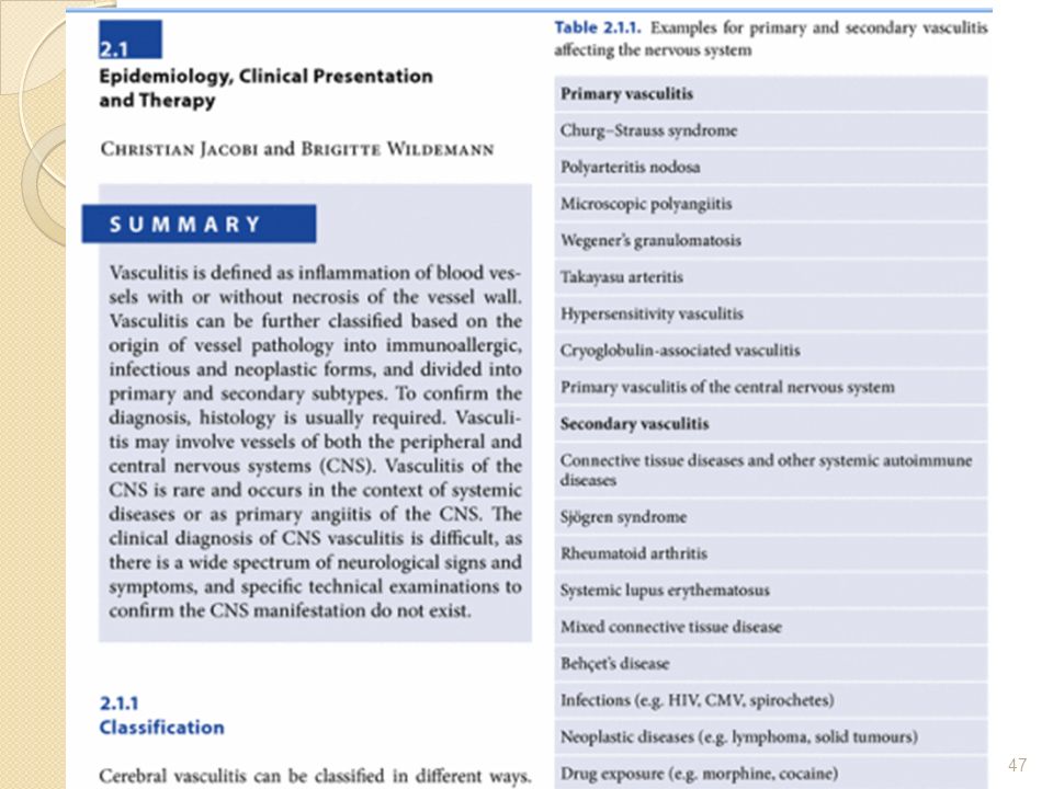
Rare cases of vasculitis have similarly been reported following rubella virus, adenovirus,echovirus, coxsackievirus, parainfluenza virus, herpes simplex viruses, and hepatitis A virus infections.

73
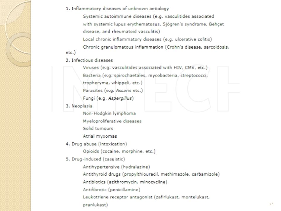
Pathology vaskulitis Perivascular cellular infiltration is a common histological finding in many disease entities, but for a definitive diagnosis of vasculitis, the presence of vascular damage, particularly in the form of fibrinoid degeneration, is necessary. Vasculitis may involve blood vessels of varying calibers and this feature forms the basis of a useful pathological classification of vasculitis. An infiltrate, composed of a variety of cell types, like neutrophils, lymphocytes, and histiocytes may invade the vessel wall and the surrounding tissue. Extravasation of red cells is a prominent feature in many vasculitides. Granulomatous inflammation with giant cell formation is a characteristic finding in some types.

74
ICHD 3

77
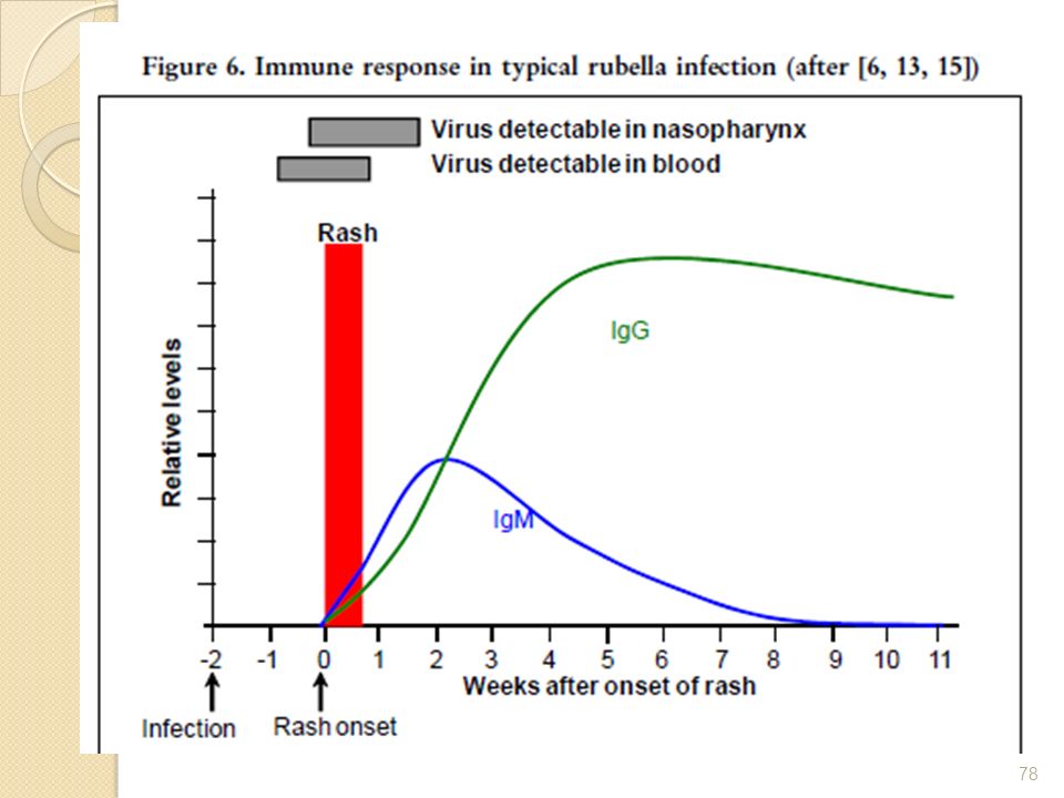
Humoral and cell-mediated immunity develop following a rubella infection. IgG and IgM antibodies are observed about days after rubella infection, at about the time when the rash appears (Figure 6). Rubella IgM antibodies wane quickly and are usually undetectable after 2 months, whereas rubella IgG antibodies persist. A rubella-specific cell-mediated lymphocyte response begins 1 week after the humoral response and persists for a lifetime. Although natural rubella infection generally confers lifelong immunity, rare cases of serologically confirmed re-infections after earlier infection (or immunization) have been reported. There have also been cases of CRS following re-infection in pregnant women with natural or vaccine-induced immunity, but this is extremely rare. Maternal rubella antibodies provide protection against rubella for the first months of life and may affect the immune response if vaccination occurs at an early age.
 have been reported. There have also been cases of CRS following re-infection in pregnant women with natural or vaccine-induced immunity, but this is extremely rare. Maternal rubella antibodies provide protection against rubella for the first months of life and may affect the immune response if vaccination occurs at an early age.")
Presentasi serupa


 Keluhan : demam RPS : Anamnesa.>")


>")


 Hidsal Jamil(135020100111028) Padel Aji Pamungkas(135020100111042)>")
